Medical Terminology Daily (MTD) is a blog sponsored by Clinical Anatomy Associates, Inc. as a service to the medical community. We post anatomical, medical or surgical terms, their meaning and usage, as well as biographical notes on anatomists, surgeons, and researchers through the ages. Be warned that some of the images used depict human anatomical specimens.

You are welcome to submit questions and suggestions using our "Contact Us" form. The information on this blog follows the terms on our "Privacy and Security Statement" and cannot be construed as medical guidance or instructions for treatment.


We have 109 guests online


A Moment in History

Jean-Louis Petit

Jean Louis Petit
(1674 – 1750)

French surgeon and anatomist, Jean Louis Petit was born in Paris in on March 13, 1674.  His family rented an apartment at his house to Alexis Littre (1658 – 1726), a French anatomist. Petit became an apprentice of Littre at seven years of age, helping him in the dissections for his lectures and at an early age became the assistant in charge of the anatomic amphitheater.

Because of Petit’s dedication to anatomy and medicine, in 1690 at the age of sixteen, became a disciple of a famous Paris surgeon, Castel.

In 1692, Petit entered the French army and performed surgery in two military campaigns. By 1693 he started delivering lectures and was accepted as a great surgeon, being invited to the most difficult operations.  In 1700 he was appointed Chief Surgeon of the Military School in Paris and in the same year he received the degree of Master of Surgery from the Faculty of Paris.

In 1715 he was made a member of the Royal Academy of Sciences and an honorary member of the Royal Society of London. He was appointed by the King as the first Director General of the Royal Academy of Surgery when it was founded in 1731.

Petit’s written works are of historical importance.  “Traite des Maladies des Os” ( A Treatise on Bone Diseases);  “Traite des Maladies Chirurgicales et des Operation” (A Treatise on Surgical Diseases and their Operations” This last book was published posthumously in 1774. He also published a monograph on hemorrhage, another on lachrymal fistula, and others.

He was one of the first to perform choIecystotomy and mastoidotomy. His original tourniquet design for amputations saved many in the battlefield and the design of the same surgical instrument today has not changed much since its invention by him.

His name is remembered in the lumbar triangle, also called the "triangle of Petit", and the abdominal hernia that can ensue through that area of weakness, the lumbar hernia or "Petit's hernia".

Sources:
1. “Jean Louis Petit – A Sketch of his Life, Character, and Writings” Hayne, AP San Fran Western Lancet 1875 4: 446-454
2. “Oeuvres compl?tes de Jean-Louis Petit” 1837 Imprimerie de F. Chapoulaud
3. Extraits de l'eloge de Jean-Louis Petit Ius dans Ia seance publique de I' Academie royale de chirurgie du 26 mai 1750” Louis A. Chirurgie 2001: 126 : 475- 81


 "Clinical Anatomy Associates, Inc., and the contributors of "Medical Terminology Daily" wish to thank all individuals who donate their bodies and tissues for the advancement of education and research”.

Click here for more information


abebooks banner

bookplateink.com


Hesselbach's triangle

Posterior view of the abdominal wall. Hesselbach's triangle
Hesselbach's triangle

Hesselbach’s triangle is a triangular region in the lower posterior aspect of the anterior abdominal wall (see yellow inset in the image). It is bound medially by the lateral border of the rectus abdominis muscle, superolaterally by the inferior (deep) epigastric vessels (label “C”) and by the inguinal ligament inferolaterally.

Hesselbach’s triangle is described as the area where a direct inguinal hernia will extrude from posterior to anterior, to protrude directly (hence the name) through the external (superficial) inguinal ring.

Franz Kaspar Hesselbach (1759-1816) was a German surgeon and anatomist who described inguinofemoral hernias in detail, publishing several books on the subject. His name is attached to several regions and structures:

• Hesselbach’s triangle, described in this article

• Hesselbach’s fascia. Known as the cribriform fascia, this perforated fascia covers the saphenous opening in the superior femoral region.

• Hesselbach’s ligament. Also known as the interfoveolar ligament, this is a thickening of the transversalis fascia in relation to the inferior (deep) epigastric vessels. 

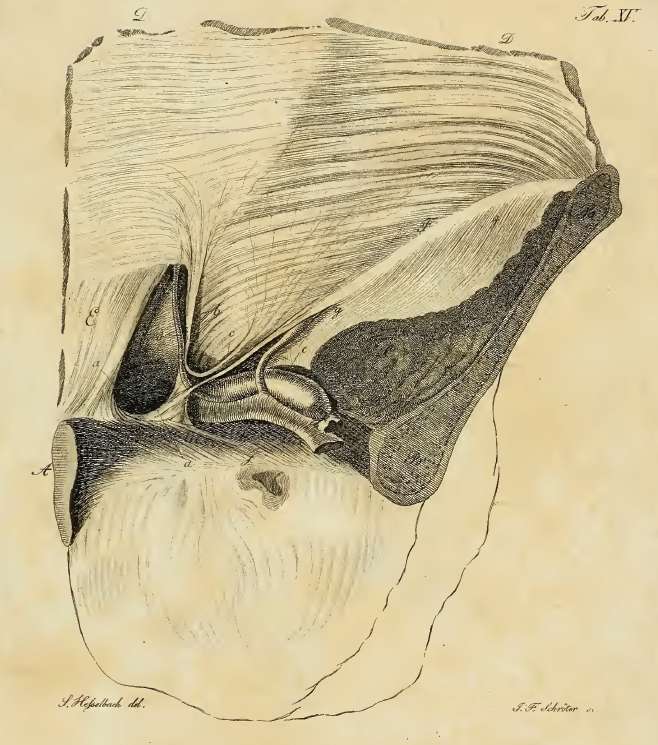
If you click on the picture, an original image by Hesselbach will appear. This image shows a defect in Hesselbach’s triangle, setting the stage for a direct inguinal hernia, as well as the interfoveolar ligament. Incidentally, Hesselbach's triangle as described today is not the area described originally by Dr. Hesselbach, where the lower border of the triangle was Cooper's ligament.

Initial image property of:CAA.Inc.Artist:M. ZuptichSecondary image by F.K. Hesselbach.


Clinical anatomy of the inguinofemoral hernias, as well as abdominal and perineal hernias are some of the lecture topics developed and delivered to the medical devices industry by Clinical Anatomy Associates, Inc. For more information Contact Us.