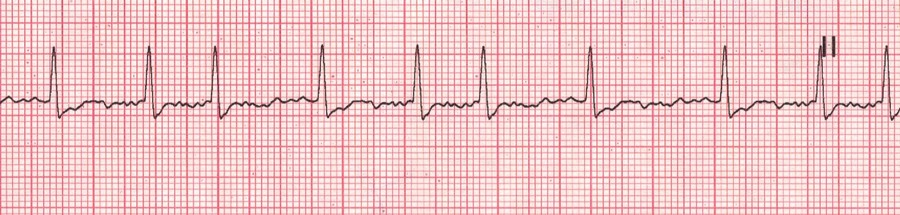
Atrial fibrillation EKG
[UPDATED] What is atrial fibrillation?
Atrial fibrillation (AFib) is one of the most common heart conditions, affecting 4% of the adult population. Characterized by a rapid, irregular heartbeat, AFib is largely due to abnormal electrical impulses that cause the atria of the heart to quiver when it should be beating steadily.The atria are the two upper chambers of the heart.
Because of this quivering action, blood flow is reduced and is not completely pumped out of the atria. This negatively impacts cardiac performance and also allows the blood to pool and potentially clot. These clots, if freed, can enter the systemic circulation and cause a stroke.
At rest, a normal heart rate is approximately 60 – 100 beats per minute. In a person with AFib, that heart rate can increase to 180 bpm or even higher. Thorough testing by your health care provider can spot abnormalities in the heart's rhythm before any obvious symptoms are noticed.
What are the symptoms?
Whether it is caused by stress, exercise, or too much caffeine, most people experience rapid heart from time to time. Most cases are harmless, but AFib is a serious medical condition that may often be long lasting. Some people with AFib experience no symptoms at all. But for others, AFib may cause:
• Exercise intolerance
• Fatigue
• Severe shortness of breath
• Chest pain
• Palpitations
• Light-headiness
What causes atrial fibrillation?
Your heart is divided into four chambers: the two upper chambers called atria, and two lower chambers called ventricles. In order for blood to be pumped through your body, a group of specialized cardiac cells, the conduction system of the heart, sends electrical impulses to the atria that tells your heart to contract. Contractions of the heart send approximately five quarts of blood through your body every minute. In people with AFib, however, the impulses are sent chaotically. The atria quiver instead of beat; the blood isn't completely pumped out and may pool and potentially clot. AFib is a leading cause of stroke because of the anatomy of the left ventricle. For more information, read this article.
Are you at risk?
Your chances of developing AFib increase with age. AFib occurs more commonly in women than in men. According to the Framingham Heart Study, AFib is associated with a higher risk of death for women than for men. You are also at greater risk of developing AFib if you suffer from an overactive thyroid, high blood pressure, a prior heart attack, congestive heart failure, valve disease, or congenital disorders.
Diagnosis
AFib can sometimes be diagnosed with a stethoscope during an exam by a doctor or other health care provider and is confirmed or diagnosed with an electrocardiogram (EKG). There are several types of EKG’s. They are:
• Resting EKG – Electrical activity in the heart is monitored when a person is at rest.
• Exercise EKG – Activity is monitored when a person jogs on a treadmill or exercises on a stationary bike.
• 24-hour EKG (Holter Monitor) – A person wears a small, portable monitor that detects activity over the course of a day.
• Transtelephonic event monitoring – A person wears a monitor for a period of a few days to several weeks. When AF is felt, the person telephones a monitoring station or activates the monitor's memory function. This type of EKG is particularly useful in detecting AF that occurs only once every few days or weeks. Unfortunately this type of monitor does not record heart events while you are sleeping.
The image on this article is a typical EKG AFib recording showing the flutter of the atria followed by the ventricular contraction. In the larger image (click on the image of the article) you can see how this fluttering of the atria causes an abnormal spacing of the ventricular contractions which some patients feel in their chest.
PERSONAL NOTE: For more information on AFib and its surgical treatment, click here.
Thanks toDr. Randall Wolf for the image andlinks