|
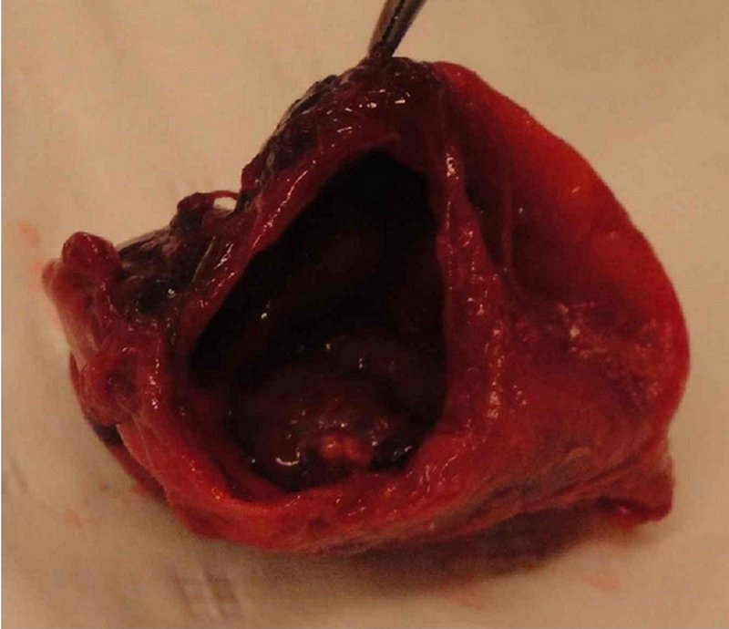
The "Canal of Nuck" is the patent embryological remnant of the processus vaginalis in the female. The processus vaginalis is an extension of the peritoneum that forms to the side of the gubernaculum, a small fibrous cord that is attached to the lower pole of the gonad in the embryo. On the other end, the gubernaculum attaches to the inner aspect of the labioscrotal fold, an embryonic structure that will become the scrotum in the male and the labia majora in the female. In the male, the processus vaginalis accompanies the gubernaculum and the testicle, on its descent towards the scrotum. In the female, the gonad (ovary) stays in the pelvis and the embryological remnants of the gubernaculum become the proper ovarian ligament (uteroovarian ligament) and the round ligament of the uterus which enters the inguinal canal, splits into multiple small fibers that disappear in the tissues of the labium majus. In the male (and female) the walls of the processus vaginalis normally fuse, closing the communication between the scrotum (and the labia majora) and the main peritoneal cavity. If they remain open, the name is different, although the pathological consequences are similar (hernia, cysts or hydrocele). In the male, it is called a “patent processus vaginalis”, and in the female it is called the “Canal of Nuck”, which is found patent in 10-20% of the cases, although its presence does not per se imply the presence of pathology. It was first described by Anton Nuck, a Dutch surgeon and anatomist (1650-1692) in his book "Adenographia & uteri anatome nova" published in 1722. In this book he questions why do some females present with inguinal hernias: "Haecce , praeter alias herniarium species , in utroque sexu obvias auditoribus meis anno fuperiori demonftrandi , difficile vifum fuit explicare , qui Hernia foeminarum inguinales orirentur?" Why when it is easy to see (the canal) in other species it is so difficult to explain to those listening why only some women have inguinal hernias? In figure XL of the same book he proceeds to show the open processus vaginalis which was from then on known as the eponymic "Canal of Nuck" The images in this article are from “Case Report: Infected Hydrocele of the Canal of Nuck” by Mandahan, P and Batthi, K. (see sources) Figure 1 shows the superficial hydrocele herniation; figure 2 shows the infected hydrocele; and figure 3 shows the excised opened hydrocele. Read the full article here. http://dx.doi.org/10.1155/2013/275257 My personal thanks to Dr. Sanford Osher who suggested this article. Dr. Miranda |
|
| Sources: 1. Adenographia curiosa et uteri foeminei anatome nova” Classic pages in Obstetrics and Gynecology Lawrence Longo, M.D. Volume 123, Issue 1, 1 September 1975, Page 66 2. Nuck, Antonio “Adenographia curiosa et uteri foeminei anatome nova” (Latin) Apud Samuellem Luchtmans https://archive.org/details/bub_gb_tQZJYL7qyssC 3. Poghosyanm T, et al “Hydrocele of canal of Nuck” Applied Radiology; Scotch Plains43.12 (Dec 2014): 37-38. 4. Bagley, J. “Cyst of Canal of Nuck.” Journal of Diagnostic Medical Sonography Vol 31, Issue 2, pp. 111 - 114 5. Bagu, A. et al Endometriosis in the canal of Nuck hydrocele: An unusual presentation 6. Mandham, P; Bhatti, K “Case Report: Infected Hydrocele of the Canal of Nuck” Case Reports in Urology Volume 2013 http://dx.doi.org/10.1155/2013/275257 |
|
| MTD Main Page | Subscribe to MTD |