Medical Terminology Daily (MTD) is a blog sponsored by Clinical Anatomy Associates, Inc. as a service to the medical community. We post anatomical, medical or surgical terms, their meaning and usage, as well as biographical notes on anatomists, surgeons, and researchers through the ages. Be warned that some of the images used depict human anatomical specimens.

You are welcome to submit questions and suggestions using our "Contact Us" form. The information on this blog follows the terms on our "Privacy and Security Statement" and cannot be construed as medical guidance or instructions for treatment.


We have 181 guests online


A Moment in History

Jean-Louis Petit

Jean Louis Petit
(1674 – 1750)

French surgeon and anatomist, Jean Louis Petit was born in Paris in on March 13, 1674.  His family rented an apartment at his house to Alexis Littre (1658 – 1726), a French anatomist. Petit became an apprentice of Littre at seven years of age, helping him in the dissections for his lectures and at an early age became the assistant in charge of the anatomic amphitheater.

Because of Petit’s dedication to anatomy and medicine, in 1690 at the age of sixteen, became a disciple of a famous Paris surgeon, Castel.

In 1692, Petit entered the French army and performed surgery in two military campaigns. By 1693 he started delivering lectures and was accepted as a great surgeon, being invited to the most difficult operations.  In 1700 he was appointed Chief Surgeon of the Military School in Paris and in the same year he received the degree of Master of Surgery from the Faculty of Paris.

In 1715 he was made a member of the Royal Academy of Sciences and an honorary member of the Royal Society of London. He was appointed by the King as the first Director General of the Royal Academy of Surgery when it was founded in 1731.

Petit’s written works are of historical importance.  “Traite des Maladies des Os” ( A Treatise on Bone Diseases);  “Traite des Maladies Chirurgicales et des Operation” (A Treatise on Surgical Diseases and their Operations” This last book was published posthumously in 1774. He also published a monograph on hemorrhage, another on lachrymal fistula, and others.

He was one of the first to perform choIecystotomy and mastoidotomy. His original tourniquet design for amputations saved many in the battlefield and the design of the same surgical instrument today has not changed much since its invention by him.

His name is remembered in the lumbar triangle, also called the "triangle of Petit", and the abdominal hernia that can ensue through that area of weakness, the lumbar hernia or "Petit's hernia".

Sources:
1. “Jean Louis Petit – A Sketch of his Life, Character, and Writings” Hayne, AP San Fran Western Lancet 1875 4: 446-454
2. “Oeuvres compl?tes de Jean-Louis Petit” 1837 Imprimerie de F. Chapoulaud
3. Extraits de l'eloge de Jean-Louis Petit Ius dans Ia seance publique de I' Academie royale de chirurgie du 26 mai 1750” Louis A. Chirurgie 2001: 126 : 475- 81


 "Clinical Anatomy Associates, Inc., and the contributors of "Medical Terminology Daily" wish to thank all individuals who donate their bodies and tissues for the advancement of education and research”.

Click here for more information


abebooks banner

bookplateink.com


Brachialis

Brachialis muscle - Image modified from the original. Public domain
Brachialis muscle.
Click on the image for a larger depiction

UPDATED: The brachialis muscle is a skeletal muscle attached proximally to the anterior surface of the humerus and distally to the coronoid process and tuberosity of the ulna. It is one of the three muscles in the anterior compartment of the arm (flexor compartment), the other two being the biceps brachii and the coracobrachialis.

It is a strong flexor of the elbow found deep to the biceps brachii. Because it does not attach to the radius, the brachialis muscle does not participate in the pronation and supination of the forearm.

The brachialis is supplied by branches of the brachial artery and by the recurrent radial artery.

The innervation of the brachialis muscle is a point to be discussed. Most modern books of anatomy state that this muscle is innervated by the musculocutaneous nerve (C5, C6, and C7). Older and more detailed books state that this muscle has a dual innervation. A 2011 research paper published in Spanish (see Sources #6) describes this dual innervation. The proximal portion of the muscles is indeed innervated by the musculocutaneous nerve, but the distal portion (in 90% of the cases) is innervated by muscular branches that arise off the radial nerve. The radial nerve (C5, C6, C7, C8 & T1) is a branch of the brachial plexus.

Following is an excerpt from the "Trail Guide to the Body" by Andrew Biel: "Ironically, (because it is deep to the biceps) the brachialis girth only helps the biceps brachii to bulge further from the arm, making the brachialis the biceps' "best friend"

Personal note: The research paper that describes the double innervation of the brachialis muscle was done at my alma mater, the University of Chile, and the authors' listing includes two of the contributors to this blog, Professors Claudio Molina and Cristian Uribe. Dr. Miranda

Sources:
1. “Gray’s Anatomy” Henry Gray, 1918
2. "Tratado de Anatomia Humana" Testut et Latarjet 8th Ed. 1931 Salvat Editores, Spain
3. "Gray's Anatomy" 42nd British Ed. Churchill Livingstone 2021
4. “An Illustrated Atlas of the Skeletal Muscles” Bowden, B. 4th Ed. Morton Publishing. 2015
5. "Trail Guide to The Body" 4th. Ed. Biel, A. Books of Discovery. 2010
6. "Doble Innervacion del Musculo Brachial en la Poblacion Chilena" Claudio Molina; Cristián Uribe; Álvaro Heras; Cristián Astorga;Jorge Lemus & Alberto Rodríguez. Int. J. Morphol, 2011. 29(4):1207-1211. A PDF copy of this paper is available here.

Note: The side image modified from the original by Anatomography, CC BY-SA 2.1 JP <https://creativecommons.org/licenses/by-sa/2.1/jp/deed.en>, via Wikimedia Commons following Creative Commons attributes.


Biceps brachii

Biceps brachii muscle - Image modified from the original, Wikimedia Commons. Public domain
Biceps brachii muscle.
Click on the image for a larger depiction

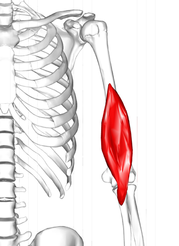
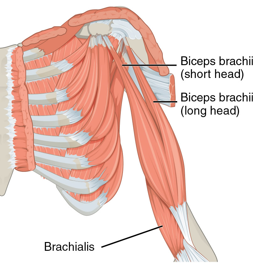
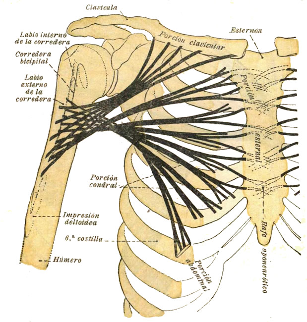
The musculus biceps brachii is a long muscle found in the anterior, aspect of the arm and is one of the three muscles contained in the anterior compartment (flexor compartment) of the arm, the other two being the brachialis and coracobrachialis muscles.  It is composed by two muscular heads, one long (lateral) , and one short (medial) that originate superiorly from separate tendons that attach to the scapula. These two heads join to  form a single long, oval-shaped belly with a single tendon that crosses the elbow joint and attaches to the radius. 

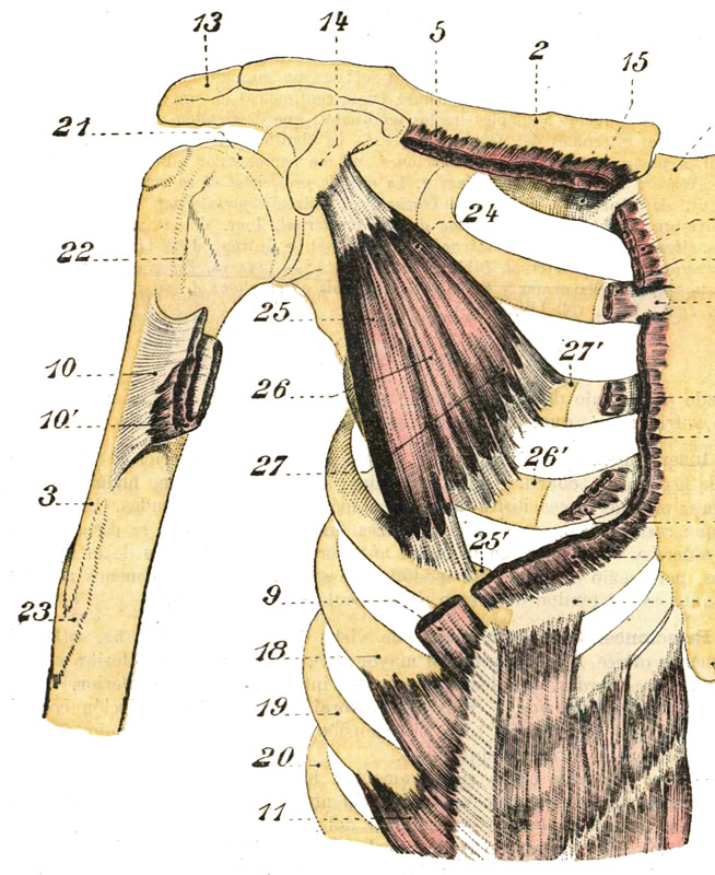
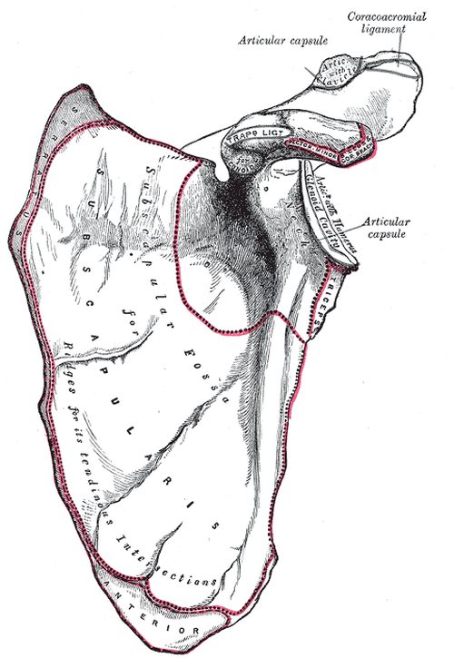
The short tendon of the biceps brachii passes anteromedial to the shoulder joint and attaches to the coracoid process of the scapula by way of a tendon that mixes with the tendon of the coracobrachialis muscle.

The long cylindrical tendon of the biceps brachii is found in the intertubercular (bicipital) groove (Lat: sulcus intertubercularis) of the humerus, and passes between the greater and lesser tubercles of the humerus, entering the articular cavity of shoulder joint, and continues superiorly to insert in the supraglenoid tubercle of the scapula.

The distal, common tendon of the biceps brachii courses inferiorly and attaches to the radial (bicipital) tuberosity of the radius. There is a well-defined bursa between the radial tuberosity and the biceps brachii tendon that allows for movement of the tendon.

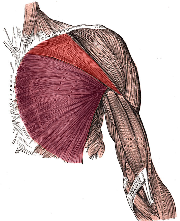
Also, a flat, fascial extension of the tendon, known as the bicipital aponeurosis extends inferomedially to blend with the antebrachial aponeurosis that covers the epitrochlear muscles of the forearm (pronator teres, flexor carpi radialis muscles). The brachial artery passes between the tendon of the biceps brachii and the bicipital aponeurosis in the anterior aspect of the elbow joint.

The biceps brachii crosses both the shoulder and the elbow join. As such, its functions will depend on which joint is fixed and which one is not. This muscle flexes the elbow, supinates the forearm, and flexes the shoulder.

It is innervated by the musculocutaneous nerve (C5,D6) which is a branch of the brachial plexus. It receives arterial supply by way of muscular branches that arise from the brachial artery.

The name of the muscle literally means "two heads" as the prefix "bi" means "two" and the Latin term "-ceps" means "head".

Note: Because the long and the short head of the biceps brachii attach to different locations of the scapula, some authors and Internet websites say that there are 18 muscles that attach to the scapula. I do not agree, as the biceps brachii is a single muscle that happens to have to separate attachments to the scapula. It would be different if this article was titled "Name the 18 separate muscular attachment points of the scapula". Dr. Miranda

The image is modified from the original via Wikimedia. Public domain. Animated image below by Wikimedia Commons - Anatomography [CC BY-SA 2.1 following Creative Commons attributes.

Niwadare, CC BY-SA 4.0 <https://creativecommons.org/licenses/by-sa/4.0>, via Wikimedia CommonsSources:
1. “Gray’s Anatomy” Henry Gray, 1918
2. "Tratado de Anatomia Humana" Testut et Latarjet 8th Ed. 1931 Salvat Editores, Spain
3. "Gray's Anatomy" 42nd British Ed. Churchill Livingstone 2021
4. “An Illustrated Atlas of the Skeletal Muscles” Bowden, B. 4th Ed. Morton Publishing. 2015
5. "Trail Guide to The Body" 4th. Ed. Biel, A. Books of Discovery. 2010


Pectoralis minor

Pectoralis minor muscle (26) - Testut & Latarjet 1931. Public domain
Pectoralis minor muscle (26)
Click on the image for a larger depiction

The pectoralis minor muscle is a small triangular muscle found deep to the pectoralis major in the anterior aspect of the thorax.

This muscle originates from three fleshy bellies that insert into the superior border and anterior surface of the third, fourth and fifth ribs. The muscle fibers converge superolaterally to insert into the inferomedial aspect of the coracoid process, of the scapula, where the tendon of the pectoralis minor intermingles and fuses with the tendon of the coracobrachialis muscle.

The pectoralis minor lies immediately anterior and covers some of the structures of the axillary region, the axillary artery and vein and some of the components of the brachial plexus. In fact, the pectoralis minor muscle is the landmark that divides the axillary artery into its three components: proximal (between the first rib and the medial border of the pectoralis minor). middle (deep to the pectoralis minor), and distal (between the lateral border of the pectoralis major and the inferior border of the teres major muscle). Thus defined the pectoralis major forms part of the anterior wall of the axilla.

In conjunction with other muscles, the pectoralis minor helps to maintain the scapular and shoulder joint in position. If the scapula is fixed, the pectoralis major assists to elevate the anterior thoracic wall during forced inhalation. The pectoralis minor also works as a depressor of the scapula and shoulder joint, abducts the scapula, and rotates the scapula.

The pectoralis minor is innervated by the medial pectoral nerve (C8.T1), a branch of the brachial plexus. Some of the fibers of the medial pectoral nerve perforate the pectoralis minor to provide nerve supply to a portion of the pectoralis major. The pectoralis minor is one of the 17 muscles that attach to the scapula.

Sources:
1. “Gray’s Anatomy” Henry Gray, 1918
2. "Tratado de Anatomia Humana" Testut et Latarjet 8th Ed. 1931 Salvat Editores, Spain
3. "Gray's Anatomy" 42nd British Ed. Churchill Livingstone 2021
4. “An Illustrated Atlas of the Skeletal Muscles” Bowden, B. 4th Ed. Morton Publishing. 2015
5. "Trail Guide to The Body" 4th. Ed. Biel, A. Books of Discovery. 2010


Pectoralis major

UPDATED: The pectoralis major muscle is the largest muscle in the anterior aspect of the thorax. It is thick and fan-shaped.  It attaches superiorly to the medial 2/3rds of the clavicle, and medially to the anterior aspect of the sternum and cartilages of the first to sixth or seventh ribs, extending inferiorly to attach to the aponeurosis of the external oblique muscle. Laterally, this muscle attaches to the lateral lip of the intertubercular groove (bicipital groove) of the humerus by a two-layered quadrilateral tendon which inserts each of the two heads of the muscle.

The superficial tendon attaches the clavicular head (red in the accompanying image), which extends between the intertubercular groove of the humerus and the clavicle. The deep tendon attaches the sternocostal head (purple in the accompanying image), which extends between the humeral intertubercular groove and the attachments in the sternum, costal cartilages, and the aponeurosis of the external oblique muscle. There is usually a well-defined interval between the two heads of the pectoralis major.

The pectoralis major is innervated by the medial pectoral nerve (C8-T1) and lateral pectoral nerve (C5-C7).

This muscle is covered by the pectoral fascia. An extension of this fascia is the clavipectoral fascia. In both male and female, the mammary gland is situated anterior to and anchors to the pectoral fascia by a number of fascial ligaments known as "Cooper's ligaments"

When both pectoral heads contract as a unit, the muscle adducts. flexes, and medially rotates the shoulder joint and humerus, such as when swimming doing and Australian crawl. Testut & Latarjet (1931) describe three separate muscular segments to this muscle, a clavicular component, a superior sternocostal component, and an inferior sternocostal component. They state that the clavicular components is quite evident, but the other two, although difficult to see, are separate. The clavicular head draws the humerus forward, upward, and medially, such as when you reach for something in front and above you. The sternocostal head draws the humerus down, forward, and medially.

The second image in this article is from Testut & Latarjet (1931) and shows the direction of muscular fibers of the three segments of the pectoralis major.

The word pectoral arises from the Latin term "pectum" meaning "chest, breast". In its true meaning, pectoral or pectoralis refers to a "chest plate" or an "adornment of the chest".

Pectoralis major muscle - Red: clavicular head. Purple: Sternocostal head - Image modified from the original by Henry VanDyke Carter, MD. Public domain
Pectoralis major muscle
Click on the image for a larger depiction 

Pectoralis major muscle - Direction of the muscular fibers. Public domain
Pectoralis major. Direction of the muscular fibers
Click on the image for a larger depiction 

Sources:
1. “Gray’s Anatomy” Henry Gray, 1918
2. "Tratado de Anatomia Humana" Testut et Latarjet 8th Ed. 1931 Salvat Editores, Spain
3. "Gray's Anatomy" 38th British Ed. Churchill Livingstone 1995
4. “An Illustrated Atlas of the Skeletal Muscles” Bowden, B. 4th Ed. Morton Publishing. 2015

First image modified from the original by Henry VanDyke Carter, MD. Public domain

Back to MTD Main Page Subscribe to MTD

Latissimus dorsi

Latissimus dorsi muscle (1) - Testut & Latarjet 1931. Public domain
Latissimus dorsi muscle
Click on the image for a larger depiction

The latissimus dorsi muscle is a large, wide, flat muscle on the posteroinferior aspect of the back. It has the shape of a triangle that has a base at the thoracolumbar spine and its apex in the axillary region.

This muscle has a wide origin by tendons that attach to the spinous processes of the lower six or seven thoracic vertebrae as well as those of the lumbar vertebrae, the sacral crest, and the posterior aspect of the external lip of the iliac crest. This created a wide fibrotendinous lamina known as the thoracolumbar fascia. The muscle also attaches to the external surface of the three or four inferiormost ribs and the inferior angle of the scapula.

From here, the muscle fibers converge superolaterally and twist anterosuperiorly to form a quadrilateral tendon that inserts deep into the bicipital groove (Lat: sulcus intertubercularis) of the humerus as shown by number 5 in the accompanying figure. There is sometimes a tendinous extension to the humeral lesser tubercle.

The latissimus dorsi extends, adducts, and medially rotates the shoulder joint, also known as the glenohumeral joint. Along with the teres major muscle they are known as the “handcuff muscles”, as this is the action of these muscles as the hands are brought together towards the back. The latissimus dorsi is innervated by the thoracodorsal (or long subscapular) nerve (C6, C7, and C8).

The Terminologia Anatomica 2 proper name is “musculus latissimus dorsi”. The plural form is “musculi latissimi dorsi”. The name of the muscle is derived from Latin. Since “latum” means “wide”, “musculus latissimus dorsi” means the “widest muscle of the back”, quite a proper name. In other languages this is more evident. In Spanish, the name for the muscle is [músculo dorsal ancho] meaning the “wide muscle of the back”.

The latissimus dorsi is one of the 17 muscles that attach to the scapula. It also forms one of the borders of the lumbar triangle of Petit, potential site for a lumbar hernia.

Sources:
1. “Gray’s Anatomy” Henry Gray, 1918
2. "Tratado de Anatomia Humana" Testut et Latarjet 8th Ed. 1931 Salvat Editores, Spain
3. "Gray's Anatomy" 42nd British Ed. Churchill Livingstone 2021
4. “An Illustrated Atlas of the Skeletal Muscles” Bowden, B. 4th Ed. Morton Publishing. 2015
5. "Trail Guide to The Body" 4th. Ed. Biel, A. Books of Discovery. 2010


Name the 17 muscles that attach to the scapula

Anterior view of the left scapula.  Image in Public Domain, by Henry Vandyke Carter, MD - Gray's Anatomy
Anterior view of the left scapula.


UPDATED:
The scapula is a flat, triangular bone that forms the posterior portion of the shoulder girdle. It is described with two surfaces, three borders, and three angles. The scapula attaches to the clavicle by way of the acromioclavicular joint and ligaments. . Seventeen muscles attach to the scapula and are listed here alphabetically:

1. Biceps brachii
2. Coracobrachialis 
3. Deltoid  
4. Infraspinatus 
5. Latissimus dorsi
6. Levator scapulae 
7. Omohyoid (inferior belly)
8. Pectoralis minor 
9. Rhomboid major 
10. Rhomboid minor 
11. Serratus anterior 
12. Subscapularis 
13. Supraspinatus 
14. Teres major 
15. Teres minor 
16. Trapezius
17. Triceps brachii (long head)

By surfaces, borders, and structures, these muscles group and attach as follows:

Posterior surface:
1. Supraspinatus
2. Infraspinatus
3. Teres major
4. Teres minor

Scapular spine and acromion:
5. Trapezius
6. Deltoid

Anterior surface:
7. Subscapularis
8. Serratus anterior

Medial border:
8. Serratus anterior
9. Rhomboid major
10. Rhomboid minor
11. Levator scapulae


Superior border:

12. Omohyoid (inferior belly)

Medial border:
13. Triceps brachii (long head)

External angle:
14. Biceps brachii (long head)

Coracoid process:
14. Biceps brachii (short head)
15. Coracobrachialis
16. Pectoralis minor

Inferior angle:

17. Latissimus dorsi

Note: Because the long and the short head of the biceps brachii attach to different locations of the scapula, some authors and Internet websites say that there are 18 muscles that attach to the scapula. I do not agree, as the biceps brachii is a single muscle that happens to have two separate attachments to the scapula. It would be different if this article was titled "Name the 18 separate muscular attachment points of the scapula". Dr. Miranda 

Sources:
1. "Tratado de Anatomia Humana" Testut et Latarjet 8 Ed. 1931 Salvat Editores, Spain
2. "Gray's Anatomy" 38th British Ed. Churchill Livingstone 1995
Image in the Public Domain, by Henry Vandyke Carter - Gray's Anatomy