|
The pelvic diaphragm is one of the four diaphragms in the human body (do you know the other three?) and it represents the lower boundary of the abdominopelvic cavity. This thin and transversely oriented structure is formed from anterior to posterior by the puboccygeus, the iliococcygeus, and the coccygeus muscles. The first two anterior muscles overlap, the pubococcygeus muscle being superior to the iliococcygeus muscle. Both of them attach laterally to a thickening of the obturator internus fascia that covers the obturator internus muscle. This thickening is known as the arcus tendineus levator ani (ATLA in the image). Because of the relation of the medial fibers of the puboccygeus muscle to the anal canal (puborectalis muscle), and what happens when these muscles contract, these two anterior muscles are known by one common name, the "levator ani" muscle. Click on the picture for a larger image. |
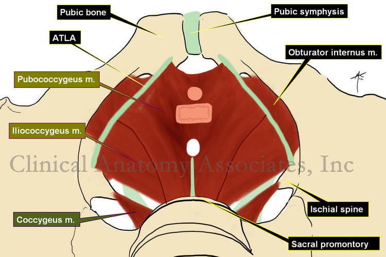 |
| The posterior component of the pelvic diaphragm is the coccygeus muscle, which is found lying on the internal aspect of the sacrospinous ligament.Image property of: CAA.Inc.. Artist: D.M. Klein. Word suggested and edited by: Dr. Sanford S. Osher, MTD Contributor | |
| Back to MTD Main Page | Subscribe to MTD |