|
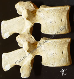
The term [intervertebral] means "between vertebrae", and [foramen] means "opening". The intervertebral foramina are bilateral openings between adjacent vertebrae. Each intervertebral foramen is found between adjacent pedicles ("P" in the large image), bound by the inferior vertebral notch and the superior vertebral notch of adjacent pedicles.
Although the term [intervertebral foramen] has been used for a long time, the concept has evolved to a more modern "intervertebral canal" or as some clinicians call it, the "lateral canal". The reason for this is that the intervertebral foramen is actually a tunnel whose length is determined by the width of the pedicles. This intervertebral canal has marked differences between the lateral, middle, and medial structures contained in the intervertebral canal.
Some of these structure are nerve roots, the dorsal root ganglion, the initial portion of the spinal nerve, dural sac, arteries, veins, recurrent nerves, fat, and a complex system of transforaminal and intraforaminal ligaments1. The structures contained in the intervertebral foramen can be compressed if the height of the intervertebral discs is compromised, or by a herniation of the intervertebral disc. The diameter of the intervertebral canal can also be reduced by bone and joint pahtology.
|
|
| If you hover over the image, the intervertebral foramen will be highlighted. For a larger version of both images, click on the legends below the image
Images property of: CAA.Inc. Photographer: D.M. Klein
1 Thoracic and lumbar intraforaminal ligaments Akdemir, G.; J Neurosurg Spine 13:351-355, 2010
|


