|
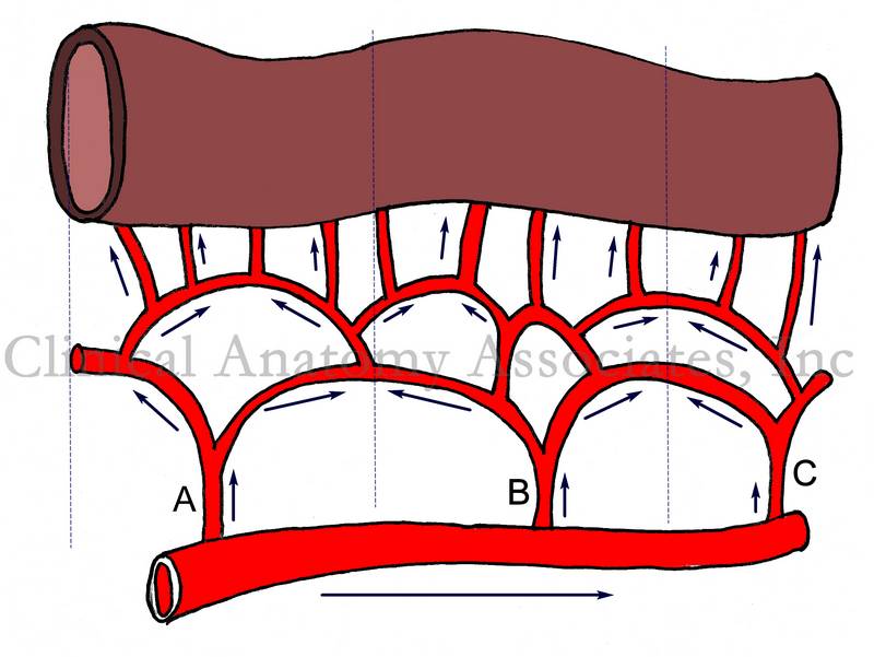
The term "collateral circulation" is generally used to denote a situation where small blood channels dilate and provide blood supply when a pathology creates a stricture and diminishes blood flow (ischemia). Although the above is correct, the term is also applicable to a normal, non-pathological situation most common in the human body. Please refer to the accompanying image for the following explanation. If needed, click on the image for a larger depiction. In the image, the arrows represent direction of flow. Most organs or organ segments receive blood supply from more than one source of blood supply. In some cases, like the stomach, there are up to four arteries that provide blood supply to the organ: the right and left gastric arteries, and the right and left gastroepiploic arteries. |
 Images property of:CAA.Inc. Artist: Dr. E. Miranda Images property of:CAA.Inc. Artist: Dr. E. Miranda |
| In other cases, like the small intestine shown in the image, blood arrives to the organ arising from several arteries (A, B, and C) that themselves arise from a parent structure. Because of hydrodynamics, the vascular territories of each artery (represented by dashed lines) tend not to overlap. If for any reason there is stenosisor blockage in any of these arteries (A,B, or C) blood will flow immediately through an alternate route and the organ will not suffer ischemia or necrosis.
This is extremely important, as these collateral channels maintain blood supply to areas that may be affected by bending, such as the elbow and knee, which have a rich collateral network. Most of the organs in the body, with some exceptions (brain, heart), have collateral circulation. Collateral circulation is extremely important for surgery, as surgeons can safely remove parts of organs without affecting the blood supply to the organ. This is also true for all gastrointestinal anastomoses. |
|
| Back to MTD Main Page | Subscribe to MTD |