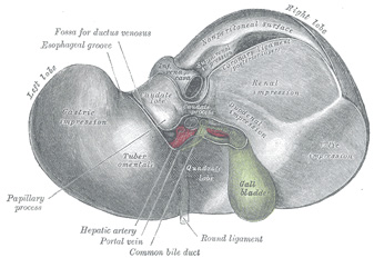
The [gallbladder] is a bile transient storage organ, part of the hepatobiliary tree, situated in the anteroinferior aspect of the liver. The gallbladder is found in a depression on the inferior aspect of the right lobe of the liver, the gallbladder fossa or fossa vesicae felleae.
In the gallbladder we describe its dome-shaped fundus, the body of the organ, and the neck which is the area that opens into the cystic duct. Close to the neck, the gallbladder has a small pouch (Hartmann's pouch) which is important for surgeons during a laparoscopic cholecystectomy, as this is where they will lock one of the instruments that allows them to manipulate the gallbladder for dissection of the organ from the gallbladder fossa (the gallbladder bed). The other surgical grasper is placed at the gallbladder fundus.
The gallbladder is composed by three layers. From deep to superficial they are:
• Mucosa: Characterized by a columnar epithelium. Towards the neck of the gallbladder the mucosa creates spiral ridges that continue in to the cystic duct.
• Fibromuscular layer: This layer is composed by connective tissue and smooth muscle, mostly longitudinal
• Serosa: This is an incomplete layer and is formed by visceral peritoneum covering the area of the gallbladder not in contact with the liver. In an unusual anatomical variation, the serosa layer can be almost complete, forming a pseudomesentery that may contains some veins.
The gallbladder receives its blood supply by way of the cystic artery, a branch of the right hepatic artery. The venous return is by way of multiple small veins that empty into the liver venous system. In some cases, these veins may form large sinuses between the liver and the gallbladder causing potential troublesome bleeding during a cholecystectomy. For those who like medical history, Dr. Eric Muhe performed the first laparoscopic cholecystectomy on September 12, 1985! We are but a few days from the 30th anniversary!
For more information on terminology on "gall-", "bile", "chol", and "chole", click here.
Sources:
1 "Tratado de Anatomia Humana" Testut et Latarjet 8 Ed. 1931 Salvat Editores, Spain
2. "Anatomy of the Human Body" Henry Gray 1918. Philadelphia: Lea & Febiger
Image modified by CAA, Inc. Original image courtesy of bartleby.com
