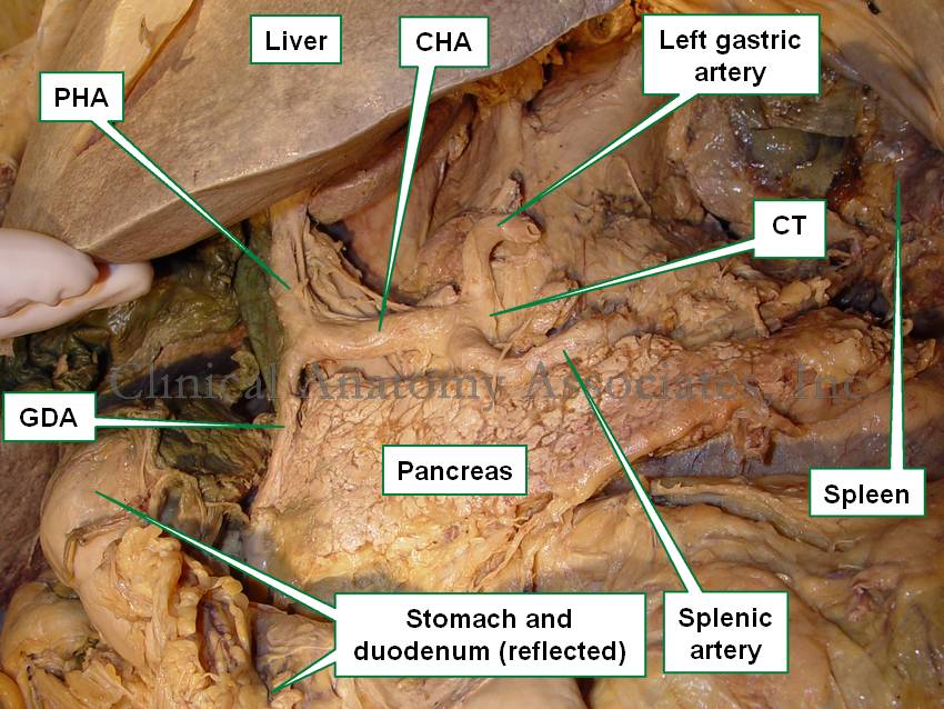
Medical Terminology Daily (MTD) is a blog sponsored by Clinical Anatomy Associates, Inc. as a service to the medical community. We post anatomical, medical or surgical terms, their meaning and usage, as well as biographical notes on anatomists, surgeons, and researchers through the ages. Be warned that some of the images used depict human anatomical specimens.

You are welcome to submit questions and suggestions using our "Contact Us" form. The information on this blog follows the terms on our "Privacy and Security Statement" and cannot be construed as medical guidance or instructions for treatment.


We have 70 guests online


A Moment in History

Jean-Louis Petit

Jean Louis Petit
(1674 – 1750)

French surgeon and anatomist, Jean Louis Petit was born in Paris in on March 13, 1674.  His family rented an apartment at his house to Alexis Littre (1658 – 1726), a French anatomist. Petit became an apprentice of Littre at seven years of age, helping him in the dissections for his lectures and at an early age became the assistant in charge of the anatomic amphitheater.

Because of Petit’s dedication to anatomy and medicine, in 1690 at the age of sixteen, became a disciple of a famous Paris surgeon, Castel.

In 1692, Petit entered the French army and performed surgery in two military campaigns. By 1693 he started delivering lectures and was accepted as a great surgeon, being invited to the most difficult operations.  In 1700 he was appointed Chief Surgeon of the Military School in Paris and in the same year he received the degree of Master of Surgery from the Faculty of Paris.

In 1715 he was made a member of the Royal Academy of Sciences and an honorary member of the Royal Society of London. He was appointed by the King as the first Director General of the Royal Academy of Surgery when it was founded in 1731.

Petit’s written works are of historical importance.  “Traite des Maladies des Os” ( A Treatise on Bone Diseases);  “Traite des Maladies Chirurgicales et des Operation” (A Treatise on Surgical Diseases and their Operations” This last book was published posthumously in 1774. He also published a monograph on hemorrhage, another on lachrymal fistula, and others.

He was one of the first to perform choIecystotomy and mastoidotomy. His original tourniquet design for amputations saved many in the battlefield and the design of the same surgical instrument today has not changed much since its invention by him.

His name is remembered in the lumbar triangle, also called the "triangle of Petit", and the abdominal hernia that can ensue through that area of weakness, the lumbar hernia or "Petit's hernia".

Sources:
1. “Jean Louis Petit – A Sketch of his Life, Character, and Writings” Hayne, AP San Fran Western Lancet 1875 4: 446-454
2. “Oeuvres compl?tes de Jean-Louis Petit” 1837 Imprimerie de F. Chapoulaud
3. Extraits de l'eloge de Jean-Louis Petit Ius dans Ia seance publique de I' Academie royale de chirurgie du 26 mai 1750” Louis A. Chirurgie 2001: 126 : 475- 81


 "Clinical Anatomy Associates, Inc., and the contributors of "Medical Terminology Daily" wish to thank all individuals who donate their bodies and tissues for the advancement of education and research”.

Click here for more information


abebooks banner

bookplateink.com


Splenic artery

The [splenic artery], also known as the [lienal artery] is one of the branches of the celiac trunk, the first unpaired branch of the abdominal aorta. Through its branches the splenic artery provides arterial blood supply to the stomach, pancreas, and spleen.

From its origin at the celiac trunk, the splenic artery goes to the left and then curves posteriorly around the spinal column. It has a very tortuous shape. In 1571 Julius Arantius (1530 -1589) described it as "tortuous, in the manner of a snake". A study by Sylvester et al (1998) measured the uncoiled (straight) lenght of the splenic artery (5.8 - 11.3 cm) as well as the variation in coiling (tortuosity) of the artery.

The splenic artery has several branches: 

Anteroinferior view of the liver and stomach, the duodenum and stomach are reflected anteriorly. CT= Celiac trunk, CHA= Common hepatic artery, PHA= Proper hepatic artery, GDA= Gastroduodenal artery
Image property of: CAA.Inc.Photographer:David M. Klein
Pancreatic arteries: Several small perforating branches. The largest of them, usually the first perforator is called either the "middle pancreatic artery",  the "great pancreatic artery", or the "arteria pancratica magna"

Left gastroepiploic artery: The largest of the splenic artery branches, this artery forms part of the greater curvature vascular arcade and provides blood supply to the left side of the stomach and part of the greater omentum

Short gastric arteries: These are several short branches that course within the gastrosplenic ligament the connects the spleen to the greater curvature and fundus of the stomach. Take down of these branches is critical in certain procedures for esophageal hiatus hernia

Splenic branches: The terminal branches of the splenic artery supply the spleen. It usually divides into a superior and an inferior branch, each one giving up to four branches that enter through the hilum of the spleen

Although rare, the splenic artery can be the site for an aneurysm. It is the third most common abominal aneurysm, after abdominal aorta and iliac artery aneurysms. They are being diagnosed more frequently now as incidental findings in cross-sectional imaging.

Sources:
1. "Tratado de Anatomia Humana" Testut et Latarjet 8 Ed. 1931 Salvat Editores, Spain
2. "Tortuosity of the Human Splenic Artery" Sylvester, PA, Stewart R, Ellis, H. Clin Anat (1996):8:214-218
3. "Splenic artery aneurysms"Trastek VF, et al. Surgery (1982) 91:694-699
9. "Splenic Artery Aneurysms and Pseudoaneurysms: Clinical Distinctions and CT Appearances" Agrawal, GA. Johnson, PT. Fishman EK. Am J Roentg (2007) 188: 4; 992-999

Back to MTD Main Page Subscribe to MTD