Medical Terminology Daily (MTD) is a blog sponsored by Clinical Anatomy Associates, Inc. as a service to the medical community. We post anatomical, medical or surgical terms, their meaning and usage, as well as biographical notes on anatomists, surgeons, and researchers through the ages. Be warned that some of the images used depict human anatomical specimens.

You are welcome to submit questions and suggestions using our "Contact Us" form. The information on this blog follows the terms on our "Privacy and Security Statement" and cannot be construed as medical guidance or instructions for treatment.


We have 177 guests online


A Moment in History

Jean-Louis Petit

Jean Louis Petit
(1674 – 1750)

French surgeon and anatomist, Jean Louis Petit was born in Paris in on March 13, 1674.  His family rented an apartment at his house to Alexis Littre (1658 – 1726), a French anatomist. Petit became an apprentice of Littre at seven years of age, helping him in the dissections for his lectures and at an early age became the assistant in charge of the anatomic amphitheater.

Because of Petit’s dedication to anatomy and medicine, in 1690 at the age of sixteen, became a disciple of a famous Paris surgeon, Castel.

In 1692, Petit entered the French army and performed surgery in two military campaigns. By 1693 he started delivering lectures and was accepted as a great surgeon, being invited to the most difficult operations.  In 1700 he was appointed Chief Surgeon of the Military School in Paris and in the same year he received the degree of Master of Surgery from the Faculty of Paris.

In 1715 he was made a member of the Royal Academy of Sciences and an honorary member of the Royal Society of London. He was appointed by the King as the first Director General of the Royal Academy of Surgery when it was founded in 1731.

Petit’s written works are of historical importance.  “Traite des Maladies des Os” ( A Treatise on Bone Diseases);  “Traite des Maladies Chirurgicales et des Operation” (A Treatise on Surgical Diseases and their Operations” This last book was published posthumously in 1774. He also published a monograph on hemorrhage, another on lachrymal fistula, and others.

He was one of the first to perform choIecystotomy and mastoidotomy. His original tourniquet design for amputations saved many in the battlefield and the design of the same surgical instrument today has not changed much since its invention by him.

His name is remembered in the lumbar triangle, also called the "triangle of Petit", and the abdominal hernia that can ensue through that area of weakness, the lumbar hernia or "Petit's hernia".

Sources:
1. “Jean Louis Petit – A Sketch of his Life, Character, and Writings” Hayne, AP San Fran Western Lancet 1875 4: 446-454
2. “Oeuvres compl?tes de Jean-Louis Petit” 1837 Imprimerie de F. Chapoulaud
3. Extraits de l'eloge de Jean-Louis Petit Ius dans Ia seance publique de I' Academie royale de chirurgie du 26 mai 1750” Louis A. Chirurgie 2001: 126 : 475- 81


 "Clinical Anatomy Associates, Inc., and the contributors of "Medical Terminology Daily" wish to thank all individuals who donate their bodies and tissues for the advancement of education and research”.

Click here for more information


abebooks banner

bookplateink.com


Mesentery

UPDATED: The term [mesentery] is of Greek origin. The prefix [mes(o)-] arises from the Greek [μέσο] meaning "middle", the root term [-enter-] means "small intestine" or "intestine", and the suffix [-y] means "process" or "structure". Thus, the mesentery is "a structure in the middle".

The term [mesentery] can be used as a generic word to denote a double-layered peritoneal membrane that stretches between an abdominal viscus and the abdominal wall. A more precise use of the term is that of mesentery proper, which extends between the posterior abdominal wall and the jejunum and ileum. The superior mesenteric artery and veins are found at the root of the mesentery proper, along with a large accumulation of lymphatic nodes, and sysmpathetic and parasympathetic nerves.

Between the two layers of the mesentery proper, there are jejunal and ileal arteries and veins, a complex system of arterial and venous arches, as well as lymphatic vessels,  autonomic nerves, and varying degrees of fat. Because of the presence of the mesentery proper, the jejunum and ileum are mobile or intraperitoneal, that is, they can slither, turn and twist with the movements of peristalsis. This movement is helped by the presence of a small amount of peritoneal fluid.

The fact that the mesentery is intraperitoneal is important in surgery. If the organ can already move around because of its mesentery, then it does not need to be "mobilized", it is already mobile!! If the organ (jejunum or ileum) have adhesions that limit their mobility within the abdominal cavity, the surgeon may have to perform and adhesiolysis to restore their mobility.

Dissection of the abdomen. The greater omentum is being pulled anteriorly

Transillumination of the transverse mesocolon. Courtesy of Michiaki Akashi, MD

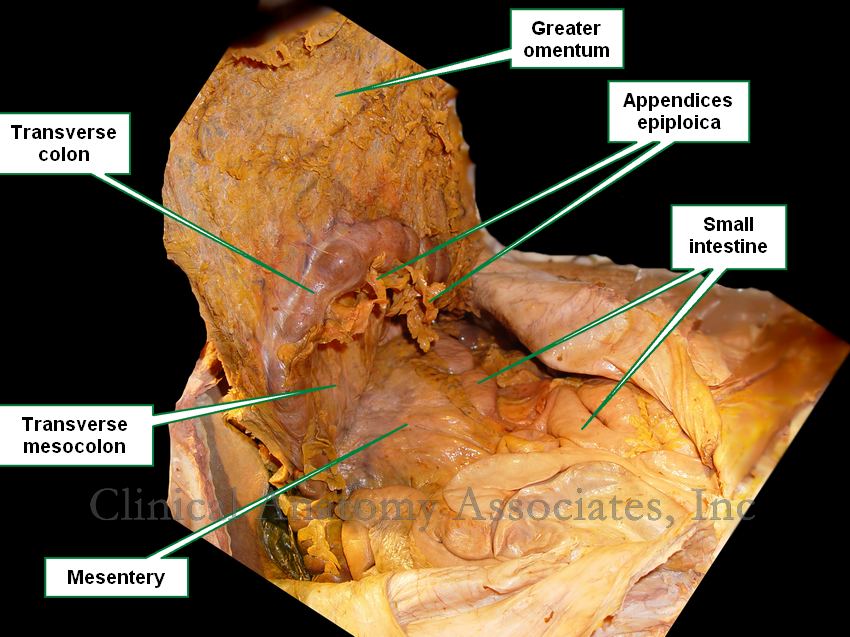
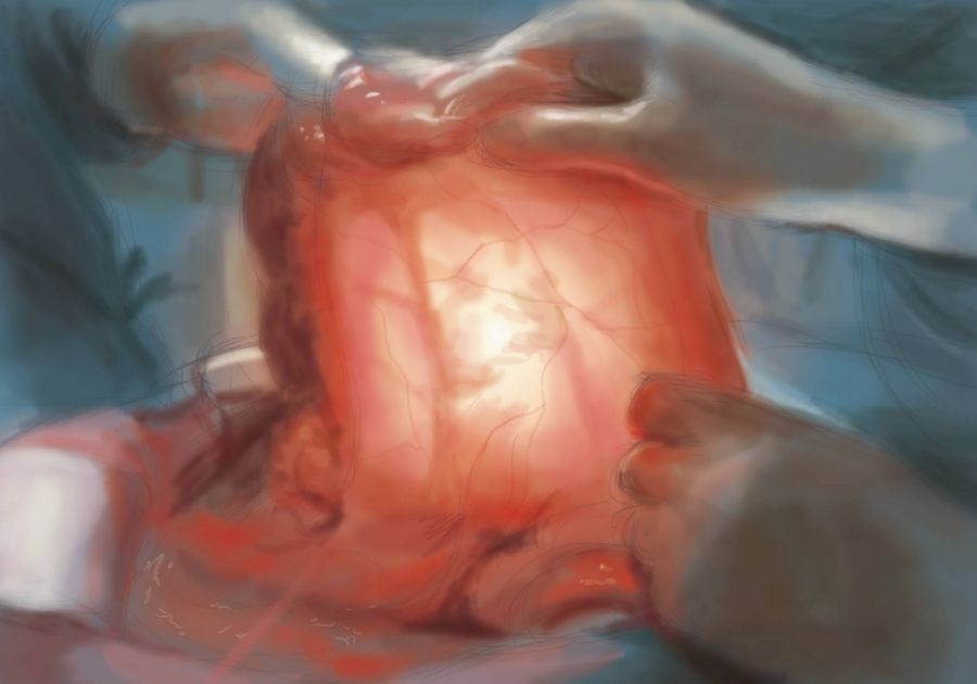
The first image shows an anatomical dissection where the greater omentum has been pulled anteriorly, exposing the small intestine and its mesentery, as well as the transverse mesocolon. Click on the image for a larger depiction. The second image (courtesy of Dr. Michiaki Akashi) is artwork depicting the surgical technique of transillumination, where the surgeon will shine a light through the mesentery to visualize the blood supply to the intestine prior to ligation and transection. The mesentery-like structure being transilluminated is the transverse mesocolon

WARNING: The first image is a photograph of a human dissection and can be considered descriptive.

NOTE: My personal thanks to Michiaki Akashi, M.D.for allowing us to use his artwork in this article. Dr. Akashi works as a surgeon and pathologist in the Saga Prefectural Hospital Koseikan in Saga, Japan.Dr. Miranda

First image property of: CAA.Inc. Photographer:David M. Klein

MTD Main Page Back to MTD Main Page