|
The sinuses of Valsalva are dilations related to both the aortic root of the ascending aorta and the root of the pulmonary trunk. These sinuses form part of the functional aspect of the corresponding aortic valve and pulmonary valve. Each one of these semilunar valves presents normally with three sinuses of Valsalva, although the sinuses of the pulmonary valve are smaller than those of the aortic valve. One of the problems encountered when describing each sinus of Valsalva is the fact that the sinus itself is not a structure, but a space. This space is found between the corresponding valve leaflet (or cusp) and the arterial wall which presents with a concavity, thus creating the sinus. This concavity is important functionally as it allows the leaflet to “flutter” in the arterial stream without getting stuck to the arterial wall. Physiological studies on the presence of the sinuses of Valsalva indicate that they play an important role decreasing of minimizing the stress of the valve leaflets. |
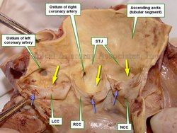
 Aortic root open. Click on the image for a larger version. |
| The dilation of the sinuses of Valsalva also creates a bulbous region at the origin of both the ascending aorta and the pulmonary trunk, the “root” of these arteries. For a better view of this bulbous region, click here. The boundary between the bulbous sinusal segment and the tubular segment of the arteries is known as the sinotubular junction (STJ).
The accompanying image shows a human ascending aorta that has been cut open to show the sinuses of Valsalva (yellow arrows), and the three cusps (leaflets) of the aortic valve. These are the non-coronary cusp (NCC), right coronary cusp (RCC), and the left coronary cusp (LCC). The ostia of the coronary arteries are visible inferior to the STJ. The sinuses of Valsalva are named after Antonio Maria Valsalva (1666 - 1723), an Italian physician and anatomist. Sources: |
|
| MTD Main Page | Back to MTD Main Page |