Medical Terminology Daily (MTD) is a blog sponsored by Clinical Anatomy Associates, Inc. as a service to the medical community. We post anatomical, medical or surgical terms, their meaning and usage, as well as biographical notes on anatomists, surgeons, and researchers through the ages. Be warned that some of the images used depict human anatomical specimens.

You are welcome to submit questions and suggestions using our "Contact Us" form. The information on this blog follows the terms on our "Privacy and Security Statement" and cannot be construed as medical guidance or instructions for treatment.


We have 249 guests online


A Moment in History

Jean-Louis Petit

Jean Louis Petit
(1674 – 1750)

French surgeon and anatomist, Jean Louis Petit was born in Paris in on March 13, 1674.  His family rented an apartment at his house to Alexis Littre (1658 – 1726), a French anatomist. Petit became an apprentice of Littre at seven years of age, helping him in the dissections for his lectures and at an early age became the assistant in charge of the anatomic amphitheater.

Because of Petit’s dedication to anatomy and medicine, in 1690 at the age of sixteen, became a disciple of a famous Paris surgeon, Castel.

In 1692, Petit entered the French army and performed surgery in two military campaigns. By 1693 he started delivering lectures and was accepted as a great surgeon, being invited to the most difficult operations.  In 1700 he was appointed Chief Surgeon of the Military School in Paris and in the same year he received the degree of Master of Surgery from the Faculty of Paris.

In 1715 he was made a member of the Royal Academy of Sciences and an honorary member of the Royal Society of London. He was appointed by the King as the first Director General of the Royal Academy of Surgery when it was founded in 1731.

Petit’s written works are of historical importance.  “Traite des Maladies des Os” ( A Treatise on Bone Diseases);  “Traite des Maladies Chirurgicales et des Operation” (A Treatise on Surgical Diseases and their Operations” This last book was published posthumously in 1774. He also published a monograph on hemorrhage, another on lachrymal fistula, and others.

He was one of the first to perform choIecystotomy and mastoidotomy. His original tourniquet design for amputations saved many in the battlefield and the design of the same surgical instrument today has not changed much since its invention by him.

His name is remembered in the lumbar triangle, also called the "triangle of Petit", and the abdominal hernia that can ensue through that area of weakness, the lumbar hernia or "Petit's hernia".

Sources:
1. “Jean Louis Petit – A Sketch of his Life, Character, and Writings” Hayne, AP San Fran Western Lancet 1875 4: 446-454
2. “Oeuvres compl?tes de Jean-Louis Petit” 1837 Imprimerie de F. Chapoulaud
3. Extraits de l'eloge de Jean-Louis Petit Ius dans Ia seance publique de I' Academie royale de chirurgie du 26 mai 1750” Louis A. Chirurgie 2001: 126 : 475- 81


 "Clinical Anatomy Associates, Inc., and the contributors of "Medical Terminology Daily" wish to thank all individuals who donate their bodies and tissues for the advancement of education and research”.

Click here for more information


abebooks banner

bookplateink.com


Scapula (2)

The [scapula] is a flat, triangular bone that forms the posterior portion of the shoulder girdle. It is described with two surfaces, three borders, and three angles. The scapula attaches to the clavicle by way of the acromioclavicular joint and ligaments. Seventeen muscles attach to the scapula providing stability and movement to the upper extremity.

The scapula has three well-defined borders. The medial border (vertebral border) is slightly convex. The superior border has a notch, the scapular notch, and a bony protuberance called the coronoid process. Where the superior and the lateral border (axillary border) meet there is a bony protuberance (the glenoid process) which has a shallow depression (the glenoid cavity), site of the glenohumeral joint or shoulder joint. Also, the lateral border presents with a small bony process just inferior to the glenoid process, the infraglenoid tubercle.

The scapula has two well-defined angles, the inferior and the superior angle, while the lateral angle is less defined because of the presence of the glenoid process.

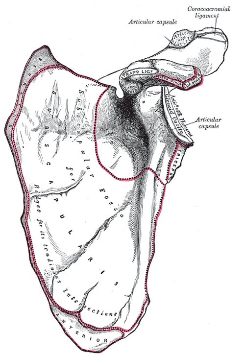
The surfaces or the scapula are the anterior and posterior surface. The anterior or costal surface is slightly concave, fairly smooth with some oblique ridges. Being concave, this area is known as the subscapular fossa. The posterior surface is separated in two by an oblique bony process call the spine of the scapula. The scapular spine ends superolaterally in a bony process called the acromion. Also, the spine of the scapula divides the posterior surface of the scapula into a supraspinous fossa and an infraspinous fossa.

Anterior view of the left scapula.  Image in Public Domain, by Henry Vandyke Carter - Gray's Anatomy
MTD Main Page Subscribe to MTD