Medical Terminology Daily (MTD) is a blog sponsored by Clinical Anatomy Associates, Inc. as a service to the medical community. We post anatomical, medical or surgical terms, their meaning and usage, as well as biographical notes on anatomists, surgeons, and researchers through the ages. Be warned that some of the images used depict human anatomical specimens.

You are welcome to submit questions and suggestions using our "Contact Us" form. The information on this blog follows the terms on our "Privacy and Security Statement" and cannot be construed as medical guidance or instructions for treatment.


We have 103 guests online


A Moment in History

Jean-Louis Petit

Jean Louis Petit
(1674 – 1750)

French surgeon and anatomist, Jean Louis Petit was born in Paris in on March 13, 1674.  His family rented an apartment at his house to Alexis Littre (1658 – 1726), a French anatomist. Petit became an apprentice of Littre at seven years of age, helping him in the dissections for his lectures and at an early age became the assistant in charge of the anatomic amphitheater.

Because of Petit’s dedication to anatomy and medicine, in 1690 at the age of sixteen, became a disciple of a famous Paris surgeon, Castel.

In 1692, Petit entered the French army and performed surgery in two military campaigns. By 1693 he started delivering lectures and was accepted as a great surgeon, being invited to the most difficult operations.  In 1700 he was appointed Chief Surgeon of the Military School in Paris and in the same year he received the degree of Master of Surgery from the Faculty of Paris.

In 1715 he was made a member of the Royal Academy of Sciences and an honorary member of the Royal Society of London. He was appointed by the King as the first Director General of the Royal Academy of Surgery when it was founded in 1731.

Petit’s written works are of historical importance.  “Traite des Maladies des Os” ( A Treatise on Bone Diseases);  “Traite des Maladies Chirurgicales et des Operation” (A Treatise on Surgical Diseases and their Operations” This last book was published posthumously in 1774. He also published a monograph on hemorrhage, another on lachrymal fistula, and others.

He was one of the first to perform choIecystotomy and mastoidotomy. His original tourniquet design for amputations saved many in the battlefield and the design of the same surgical instrument today has not changed much since its invention by him.

His name is remembered in the lumbar triangle, also called the "triangle of Petit", and the abdominal hernia that can ensue through that area of weakness, the lumbar hernia or "Petit's hernia".

Sources:
1. “Jean Louis Petit – A Sketch of his Life, Character, and Writings” Hayne, AP San Fran Western Lancet 1875 4: 446-454
2. “Oeuvres compl?tes de Jean-Louis Petit” 1837 Imprimerie de F. Chapoulaud
3. Extraits de l'eloge de Jean-Louis Petit Ius dans Ia seance publique de I' Academie royale de chirurgie du 26 mai 1750” Louis A. Chirurgie 2001: 126 : 475- 81


 "Clinical Anatomy Associates, Inc., and the contributors of "Medical Terminology Daily" wish to thank all individuals who donate their bodies and tissues for the advancement of education and research”.

Click here for more information


abebooks banner

bookplateink.com


Valve of the inferior vena cava

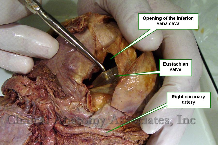
The [valve of the inferior vena cava] is probably better known by its eponym, the [Eustachian valve]. This is an incomplete valve found at the most distal end of the inferior vena cava, at the point where it opens into the right atrium of the heart. The valve is muscular at its base and composed mostly by membranous endocardium, and is normally the only valve present in the inferior vena cava.

It appears as a membranous semilunar fold extending posteriorly from the limbus fossa ovalis, anterior to the inferior caval orifice where it disappears. Its free border is usually membranous, concave, and directed anterosuperiorly. Its  base is a transverse muscular ridge continuous with the anterior margin of the caval orifice.

It has no function in the adult, but in the fetus it helps to divert the flow of blood coming from the inferior vena cava towards the foramen ovale [fossa ovalis], as part of fetal circulation.

Eustachian valve
Image property of CAA, Inc.
Click on the image for a larger depiction

It has great anatomical variation in size, shape and thickness. On its medial aspect it joins with the Thebesian valve at the orifice of the coronary sinus.

First described by Bartolomeo Eustachius (c1500 - 1574) in 1653, the existence of the valve of the inferior vena cava was disputed by many until Winslow in 1717 confirmed its presence and suggested its function in fetal circulation.

Sources:
1. "The Valve of the Inferior Vena Cava" Hickie, JB Br Heart J. 1956 Jul; 18(3): 320–326
2. "Bartolommeo Eustachio; a great medical genius whose masterpiece remained hidden for 150 years" Wells, WA Arch Otolaring (1925) 48: 58

MTD Main Page Subscribe to MTD