|
The temporal muscle (Lat:Temporalis) is a bilateral muscle located on the side of the head. It belongs to a subgroup of head muscles called Masticatory Muscles, named after their function elevating the mandible to produce the mandible movements (1,2). Masticatory muscles are four per side: Temporalis, Masseter, Pterygoideus medialis and Pterygoideus lateralis (1,2).
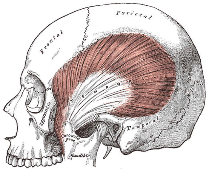
The temporalis muscle is a fan-shaped muscle which occupies the temporal fossa from which its fascicles (fibers) converge to the coronoid process of the mandible. Classic description for this muscle recognizes three main muscular bodies (anterior, midle, and posterior) originated from the temporal fossa up to the lower temporalis line and the temporalis fascia, fascicles which descend through the inner part de the zygomatic arch converging to be inserted on the coronoid process of the mandible, its temporalis crest and anterior margin of the mandibular branch through thick tendons (1,2).
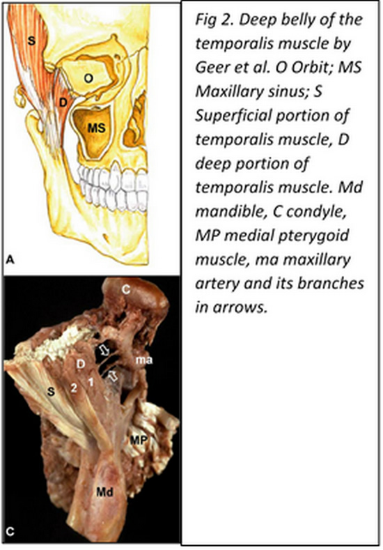
In 1996 Dunn et al. (3) reported the discovery of a so far unknown masticatory muscle called the “sphenomandibularis”, originated from the greater wing of the sphenoid bone medial to the temporalis muscle and descends on an oblique (lateral and slightly posterior) fashion reaching distally the coronoid process of the mandible. This muscular portion has been recognized as the “deep belly of the temporalis muscle” and has been described by several authors since then (4,5,6,8). The importance that has been given to this particular bundle lies on the fact that its medial insertion can reach a close relationship to the foramen rotundum, place of emergency from the cranium of the maxillary nerve, which has been hypothesized, could lead to eventual alteration of this nerve if it got trapped by this part of the muscle (6, 7).
The temporalis muscle receives innervation fundamentally from branches of the mandibular nerve: Deep temporal nerve (N. temporalis profundus)through its anterior middle and posterior branches.
The temporalis muscle is covered by a thick fascia layer: the temporalis fascia.
Article written by: Maria F. Cortés, DDS, MSc.
Images from:
Fig 1. Public domain, by Henry Vandyke Carter, MD - Gray's Anatomy, 1918
Fig 2. Geers C, Nyssen-Behets C, Cosnard G, Lengelé B. The deep belly of the temporalis muscle: an anatomical, histological and MRI study. Surg Radiol Anat. 2005 Aug;27(3):184-91. Epub 2005 Apr 9
|
Click on the image for a larger depiction
|
Sources:
1. “Anatomía humana” V.2. Latarjet- Ruiz Liard, 4ª ed. 6ª reimp. 2008 Médica Panamericana, Buenos Aires, Argentina.
2. “Anatomía humana: descriptiva, topográfica y funcional. Tomo 1. Cabeza y Cuello, Rouviere H – Delmas A, 11° ed. 2005 MASSON, S.A., Barcelona, Spain.
3. Dunn GF, Hack GD, Robinson WL, Koritzer RT. Anatomical observation of a craniomandibular muscle originating from the skull base: the sphenomandibularis. Cranio. 1996 Apr;14(2):97-103; discussion 104-5.
4. Shimokawa T, Akita K, Soma K, Sato T. Innervation analysis of the small muscle bundles attached to the temporalis: truly new muscles or merely derivatives of the temporalis? Surg Radiol Anat. 1998;20(5):329-34.
5. Akita K, Shimokawa T, Sato T. Aberrant muscle between the temporalis and the lateral pterygoid muscles: M. pterygoideus proprius (Henle). Clin Anat. 2001 Jul;14(4):288-91.
6. Schön Ybarra MA, Bauer B. Medial portion of M. Temporalis and its potential involvement in facial pain. Clin Anat. 2001;14(1):25-30.
7. Fuentes E, Llanos S, Gómez R, Llanos P, Llanos F, Cortés-Sylvester MF, Solaria P, Melian A, Asfura J, Santos M, Zamorano E. Discovery of deep temporalis muscle belly close to maxillary nerve in a patient with trigeminal neuralgia: hypothesis of muscular compression and case report treated by Botox® Onabotulinum toxin tipe-A. Chirurgia 2016 June;29(3):99-102
8. Geers C, Nyssen-Behets C, Cosnard G, Lengelé B. The deep belly of the temporalis muscle: an anatomical, histological and MRI study. Surg Radiol Anat. 2005 Aug;27(3):184-91. Epub 2005 Apr 9. |