Sponsor   

Medical Terminology Daily (MTD) is a blog sponsored by Clinical Anatomy Associates, Inc. as a service to the medical community, medical students, and the medical industry. We post anatomical, medical or surgical terms, their meaning and usage, as well as biographical notes on anatomists, surgeons, and researchers through the ages. Be warned that some of the images used depict human anatomical specimens.

Click on the link below to subscribe to the MTD newsletter. If you think an article could be interesting to somebody else, feel free to forward the link of the article. Should you want to use the information on the article, please follow the CAA, Inc Privacy and Security Statement found at the bottom of this page. 

You are welcome to submit questions and suggestions using our "Contact Us" form. The information on this blog follows the terms on our "Privacy and Security Statement"  and cannot be construed as medical guidance or instructions for treatment. 


We have 145 guests online


A Moment in History

Self-portrait, Henry Vandyke Carter, MD (Public Domain)

Henry Vandyke Carter, MD
(1831 – 1897)

English physician, surgeon, medical artist, and a pioneer in leprosy and mycetoma studies.  HV Carter was born in Yorkshire in 1831. He was the son of Henry Barlow Carter, a well-known artist and it is possible that he honed his natural talents with his father. His mother picked his middle name after a famous painter, Anthony Van Dyck. This is probably why his name is sometimes shown as Henry Van Dyke Carter, although the most common presentation of his middle name is Vandyke.

Having problems to finance his medical studies, HV Carter trained as an apothecary and later as an anatomical demonstrator at St. George’s Hospital in London, where he met Henry Gray (1872-1861), who was at the time the anatomical lecturer. Having seen the quality of HV Carter’s drawings, Henry Gray teamed with him to produce one of the most popular and longer-lived anatomy books in history: “Gray’s Anatomy”, which was first published in late 1857.  The book itself, about which many papers have been written, was immediately accepted and praised because of the clarity of the text as well as the incredible drawings of Henry Vandyke Carter.

While working on the book’s drawings, HV Carter continued his studies and received his MD in 1856.

In spite of initially being offered a co-authorship of the book, Dr. Carter was relegated to the position of illustrator by Henry Gray and never saw the royalties that the book could have generated for him. For all his work and dedication, Dr. Carter only received a one-time payment of 150 pounds. Dr.  Carter never worked again with Gray, who died of smallpox only a few years later.

Frustrated, Dr. Carter took the exams for the India Medical Service.  In 1858 he joined as an Assistant Surgeon and later became a professor of anatomy and physiology. Even later he served as a Civil Surgeon. During his tenure with the India Medical Service he attained the ranks of Surgeon, Surgeon-Major, Surgeon-Lieutenant-Colonel, and Brigade-Surgeon.

Dr. Carter dedicated the rest of his life to the study of leprosy, and other ailments typical of India at that time. He held several important offices, including that of Dean of the Medical School of the University of Bombay. In 1890, after his retirement, he was appointed Honorary Physician to the Queen.

Dr. Henry Vandyke Carter died of tuberculosis in 1897.

Personal note: Had history been different, this famous book would have been called “Gray and Carter’s Anatomy” and Dr. Carter never gone to India. His legacy is still seen in the images of the thousands of copies of “Gray’s Anatomy” throughout the world and the many reproductions of his work available on the Internet. We are proud to use some of his images in this blog. The image accompanying this article is a self-portrait of Dr. Carter. Click on the image for a larger depiction. Dr. Miranda

Sources:
1. “Obituary: Henry Vandyke Carter” Br Med J (1897);1:1256-7
2. “The Anatomist: A True Story of ‘Gray’s Anatomy” Hayes W. (2007) USA: Ballantine
3. “A Glimpse of Our Past: Henry Gray’s Anatomy” Pearce, JMS. J Clin Anat (2009) 22:291–295
4. “Henry Gray and Henry Vandyke Carter: Creators of a famous textbook” Roberts S. J Med Biogr (2000) 8:206–212.
5. “Henry Vandyke Carter and his meritorious works in India” Tappa, DM et al. Indian J Dermatol Venereol Leprol (2011) 77:101-3


"Clinical Anatomy Associates, Inc., and the contributors of "Medical Terminology Daily" wish to thank all individuals who donate their bodies and tissues for the advancement of education and research”.

Click here for more information


Rare & Collectible Books at AbeBooks.com 

bookplateink.com


Coumadin ridge

UPDATED: The [Coumadin ridge], also known as the [Warfarin ridge], or a [left atrial pseudotumor]. is an excessive elevation or protrusion of a normal ridge found between the left superior pulmonary vein and the internal ostium of the left atrial appendage. Usually this ridge will extend inferiorly towards and anterior to the ostium of the left inferior pulmonary vein. The Coumadin ridge is considered an anatomical variation of the otherwise small ridge, known as the left lateral ridge.

Because of its location and morphology, some cardiologists and radiologists have mistaken this elevation or fold of the internal anatomy of the left atrium for a thrombus and prescribed anticoagulant therapy (Coumadin or Warfarin) when none was needed, hence its name.

Coumadin ridge
Click on the image for a larger version
To understand the generation of the Coumadin ridge we must understand the embryology of this area of the heart. The left atrial appendage is the original left atrium in the embryo, which is displaced anteriorly and superolaterally when the veins that enter the atrium start to dilate at their distal end creating the left sinus venarum. After the left atrium proper has formed, the left atrial appendage is left as nothing more than an embryological remnant that can cause problems if the patient has atrial fibrillation (AFib). The ridge forms at the point where the left atrial appendage and the sinus venarum meet.

The Coumadin ridge can vary in morphology, from presenting as an elevated ridge, to a bulbous, pedunculated mass that seems to float within the left atrial appendage and undulate, following the cardiac motion, forcing the cardiologist into believing they are in the presence of a thrombus or a tumor within the heart.

This fold of tissue may contain the ligament of Marshall, autonomic nerves, and a small artery. In rare cases there may be an actual tumor arising from the location of the Coumadin ridge, but this is just a coincidence.

Now that the Coumadin ridge is a better known anatomical variation, cardiologist sometimes refer to their finding as a pseudotumor, a description that may scare the patient, but is only but a fold of tissue inside the heart.

Finding a Coumadin ridge in a patient with atrial fibrillation can be an interesting situation requiring differential diagnosis, as a patient with AFib can present with thrombi in the left atrial appendage. What to do? Is it or is it not a thrombus? Also, a differential diagnosis is needed in the case where the image is actually that of a left atrial tumor or an atrial myxoma.

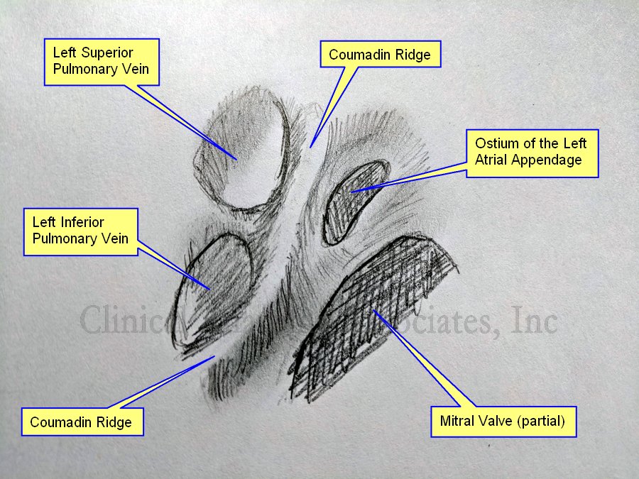
The accompanying image is own work based on Sra (2004) and McKay (2008), and is a graphite on paper sketch. The image shown an internal view of the left atrium showing the left superior and inferior pulmonary vein, the ostium of the left atrial appendage and a segment of the area of the mitral valve.

We would like to thank Dr. Randall K Wolf, a contributor to Medical Terminology Daily for suggesting this article.

Sources:
1. “Coumadin ridge: An incidental finding of a left atrial pseudotumor on transthoracic echocardiography” Lohdi,AM, et al. World J Clin Cases. 2015 Sep 16; 3(9): 831–834
2. “Coumadin ridge” Tasco, V. https://radiopaedia.org/articles/coumadin-ridge
3. “Papillary fibroelastoma arising from the coumadin ridge” Malik, M, Shilo, K, Kilic,A. J Cardiovasc Thorac Res. 2017;9(2):118-120.
4. “‘Coumadin ridge’ in the left atrium demonstrated on three dimensional transthoracic echocardiography” McKay,T., Thomas, L. Europ J Echocard (2008) 9, 298–300
5. “Endocardial imaging of the left atrium in patients with atrial fibrillation” Sra J; Krum D; Okerlund D; Thompson H. J Cardiovasc Electrophysiol 2004 Feb; Vol. 15 (2), pp. 247