Sponsor   

Medical Terminology Daily (MTD) is a blog sponsored by Clinical Anatomy Associates, Inc. as a service to the medical community, medical students, and the medical industry. We post anatomical, medical or surgical terms, their meaning and usage, as well as biographical notes on anatomists, surgeons, and researchers through the ages. Be warned that some of the images used depict human anatomical specimens.

Click on the link below to subscribe to the MTD newsletter. If you think an article could be interesting to somebody else, feel free to forward the link of the article. Should you want to use the information on the article, please follow the CAA, Inc Privacy and Security Statement found at the bottom of this page. 

You are welcome to submit questions and suggestions using our "Contact Us" form. The information on this blog follows the terms on our "Privacy and Security Statement"  and cannot be construed as medical guidance or instructions for treatment. 


We have 161 guests online


A Moment in History

Self-portrait, Henry Vandyke Carter, MD (Public Domain)

Henry Vandyke Carter, MD
(1831 – 1897)

English physician, surgeon, medical artist, and a pioneer in leprosy and mycetoma studies.  HV Carter was born in Yorkshire in 1831. He was the son of Henry Barlow Carter, a well-known artist and it is possible that he honed his natural talents with his father. His mother picked his middle name after a famous painter, Anthony Van Dyck. This is probably why his name is sometimes shown as Henry Van Dyke Carter, although the most common presentation of his middle name is Vandyke.

Having problems to finance his medical studies, HV Carter trained as an apothecary and later as an anatomical demonstrator at St. George’s Hospital in London, where he met Henry Gray (1872-1861), who was at the time the anatomical lecturer. Having seen the quality of HV Carter’s drawings, Henry Gray teamed with him to produce one of the most popular and longer-lived anatomy books in history: “Gray’s Anatomy”, which was first published in late 1857.  The book itself, about which many papers have been written, was immediately accepted and praised because of the clarity of the text as well as the incredible drawings of Henry Vandyke Carter.

While working on the book’s drawings, HV Carter continued his studies and received his MD in 1856.

In spite of initially being offered a co-authorship of the book, Dr. Carter was relegated to the position of illustrator by Henry Gray and never saw the royalties that the book could have generated for him. For all his work and dedication, Dr. Carter only received a one-time payment of 150 pounds. Dr.  Carter never worked again with Gray, who died of smallpox only a few years later.

Frustrated, Dr. Carter took the exams for the India Medical Service.  In 1858 he joined as an Assistant Surgeon and later became a professor of anatomy and physiology. Even later he served as a Civil Surgeon. During his tenure with the India Medical Service he attained the ranks of Surgeon, Surgeon-Major, Surgeon-Lieutenant-Colonel, and Brigade-Surgeon.

Dr. Carter dedicated the rest of his life to the study of leprosy, and other ailments typical of India at that time. He held several important offices, including that of Dean of the Medical School of the University of Bombay. In 1890, after his retirement, he was appointed Honorary Physician to the Queen.

Dr. Henry Vandyke Carter died of tuberculosis in 1897.

Personal note: Had history been different, this famous book would have been called “Gray and Carter’s Anatomy” and Dr. Carter never gone to India. His legacy is still seen in the images of the thousands of copies of “Gray’s Anatomy” throughout the world and the many reproductions of his work available on the Internet. We are proud to use some of his images in this blog. The image accompanying this article is a self-portrait of Dr. Carter. Click on the image for a larger depiction. Dr. Miranda

Sources:
1. “Obituary: Henry Vandyke Carter” Br Med J (1897);1:1256-7
2. “The Anatomist: A True Story of ‘Gray’s Anatomy” Hayes W. (2007) USA: Ballantine
3. “A Glimpse of Our Past: Henry Gray’s Anatomy” Pearce, JMS. J Clin Anat (2009) 22:291–295
4. “Henry Gray and Henry Vandyke Carter: Creators of a famous textbook” Roberts S. J Med Biogr (2000) 8:206–212.
5. “Henry Vandyke Carter and his meritorious works in India” Tappa, DM et al. Indian J Dermatol Venereol Leprol (2011) 77:101-3


"Clinical Anatomy Associates, Inc., and the contributors of "Medical Terminology Daily" wish to thank all individuals who donate their bodies and tissues for the advancement of education and research”.

Click here for more information


Rare & Collectible Books at AbeBooks.com 

bookplateink.com


Prevention of Stroke in Atrial Fibrillation - Elimination of the Left Atrial Appendage


If you arrived to this website looking for information on Atrial Fibrillation, you will find some here and in this article.


Prevention of Stroke in Atrial Fibrillation;
Elimination of the Left Atrial Appendage.
An online educational video.

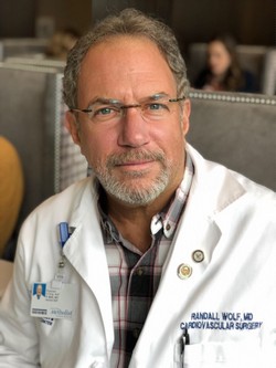
Randall, K. Wolf MD, FACS, FACC


This video educational program is hosted by the Houston Methodist DeBakey Heart and Vascular Center Education Center

Atrial fibrillation (AFib) is estimated to affect up to 4% of the population. Characterized by a rapid, irregular heartbeat, AFib is largely due to abnormal electrical impulses that cause the atria of the heart to quiver instead of beating steadily. Blood flow is reduced and is not completely pumped out of the two small upper chambers of the heart, the atria. This negatively impacts cardiac performance and also allows the blood to pool and potentially clot, especially in an extension of the left atrium, the left atrial appendage (LAA). At rest, a normal heart rate is approximately 60 – 100 beats per minute. In a person with AFib, that heart rate can increase to 180 bpm or even higher.

rwolfmf sm
The main concern with AFib and stagnant blood flow in the LAA is the potential for clot formation (thrombus). While this can happen in either atria (right or left) the anatomy of the right atrium and right atrial appendage are less conducive to clot formation. The LAA is exactly the opposite and the clots, should they float into the bloodstream, tend to enter the larger arteries that go towards the head and brain, increasing the chances for a stroke.

In this educational video Dr. Wolf discusses the above, as well as the benefits of the elimination of the LAA, decreasing blood pressure, decreasing the chances of a stroke, and helping return the heart to normal rhythm.

Dr. Wolf is a surgical innovator who since the year 2000 has been a pioneer in the minimally invasive surgical treatment of AFib. He has performed over 2000 Wolf MiniMaze procedures since the first one in 2003 and has demonstrated the procedure to over 800 heart surgeons worldwide. He has been visiting professor in 18 countries, including Oxford University, University of Tokyo and Peking University. Dr. Wolf has delivered hundreds of invited lectures at hospitals, academic meetings and seminars in the United States and abroad.

Dr. Wolf is currently a member of the DeBakey Heart and Vascular Center at Houston Methodist Hospital in the Texas Medical Center. He serves as the arrhythmia specialist of the group. In 2018 Dr. Wolf operated on AF patients from 32 US states. He was also the keynote speaker at the annual Japanese Society for Tobacco Control in Takamatsu, Japan and the annual Chinese Society of Cardiothoracic Surgeons in Shenyang, China.


videocover011  When you click the above image you will be taken to the YouTube DeBakey Education Channel
in a separate window

NOTE: Dr. Randall Wolf is a contributor to Clinical Anatomy Associates. My personal thanks to him and to the DeBakey Heart and Vascular Center for their invitation to participate in this educational video. Dr. Miranda.