Medical Terminology Daily (MTD) is a blog sponsored by Clinical Anatomy Associates, Inc. as a service to the medical community. We post anatomical, medical or surgical terms, their meaning and usage, as well as biographical notes on anatomists, surgeons, and researchers through the ages. Be warned that some of the images used depict human anatomical specimens.

You are welcome to submit questions and suggestions using our "Contact Us" form. The information on this blog follows the terms on our "Privacy and Security Statement" and cannot be construed as medical guidance or instructions for treatment.


We have 148 guests online


A Moment in History

Jean-Louis Petit

Jean Louis Petit
(1674 – 1750)

French surgeon and anatomist, Jean Louis Petit was born in Paris in on March 13, 1674.  His family rented an apartment at his house to Alexis Littre (1658 – 1726), a French anatomist. Petit became an apprentice of Littre at seven years of age, helping him in the dissections for his lectures and at an early age became the assistant in charge of the anatomic amphitheater.

Because of Petit’s dedication to anatomy and medicine, in 1690 at the age of sixteen, became a disciple of a famous Paris surgeon, Castel.

In 1692, Petit entered the French army and performed surgery in two military campaigns. By 1693 he started delivering lectures and was accepted as a great surgeon, being invited to the most difficult operations.  In 1700 he was appointed Chief Surgeon of the Military School in Paris and in the same year he received the degree of Master of Surgery from the Faculty of Paris.

In 1715 he was made a member of the Royal Academy of Sciences and an honorary member of the Royal Society of London. He was appointed by the King as the first Director General of the Royal Academy of Surgery when it was founded in 1731.

Petit’s written works are of historical importance.  “Traite des Maladies des Os” ( A Treatise on Bone Diseases);  “Traite des Maladies Chirurgicales et des Operation” (A Treatise on Surgical Diseases and their Operations” This last book was published posthumously in 1774. He also published a monograph on hemorrhage, another on lachrymal fistula, and others.

He was one of the first to perform choIecystotomy and mastoidotomy. His original tourniquet design for amputations saved many in the battlefield and the design of the same surgical instrument today has not changed much since its invention by him.

His name is remembered in the lumbar triangle, also called the "triangle of Petit", and the abdominal hernia that can ensue through that area of weakness, the lumbar hernia or "Petit's hernia".

Sources:
1. “Jean Louis Petit – A Sketch of his Life, Character, and Writings” Hayne, AP San Fran Western Lancet 1875 4: 446-454
2. “Oeuvres compl?tes de Jean-Louis Petit” 1837 Imprimerie de F. Chapoulaud
3. Extraits de l'eloge de Jean-Louis Petit Ius dans Ia seance publique de I' Academie royale de chirurgie du 26 mai 1750” Louis A. Chirurgie 2001: 126 : 475- 81


 "Clinical Anatomy Associates, Inc., and the contributors of "Medical Terminology Daily" wish to thank all individuals who donate their bodies and tissues for the advancement of education and research”.

Click here for more information


abebooks banner

bookplateink.com


Septum

The term [septum] is Latin, and means "wall", "division", or "partition". The plural form for [septum] is [septa]. Examples of use of this word are:

• Nasal septum: The osteocartilaginous partition between both sides of the nose
• Septum pellucidum: A transparent division between the lateral ventricles of the brain.
• Interatrial septum: The thin, muscular separation between the cardiac atria (see image, item "E")
• Interventricular septum: The muscular and membranous division between the ventricles of the heart (see image, item "D")

Image property of: CAA.Inc.. Photographer: D.M. Klein. 

Heart model - LAO cranial view
Back to MTD Main Page Back to MTD Main Page

Arachnoid mater

The arachnoid mater, commonly referred to only as [arachnoid], is one of the three meninges that surround, cover, and protect the spinal cord and the brain. 

The first word is Greek, from [arachne] meaning "spider" and the suffix [-oid], meaning "similar to". The second word is Latin, from [mater], meaning "mother". The name arachnoid refers to the fact that in a fresh specimen the arachnoid mater looks quite like a spider's web. See the accompanying image. Click on the image for a larger version.

Embryologically, the arachnoid mater and the pia mater are considered to have a common origin, and are called the leptomeninges (thin meninges). These two membranes, internal to the dura mater, are very thin and separated by the subarachnoid space, which is filled with cerebrospinal fluid

Spinal cord dissection, posterior viewImage property of: CAA.Inc.
Photographer: D.M. Klein

The cerebrospinal fluid (CSF) is produced by the choroid plexuses in the ventricular system of the brain, to then pass externally into the subarachnoid space. Excess production of CSF is one of the causes of hydrocephalus.
 
Back to MTD Main Page Subscribe to MTD

Thorax

The word [thorax] is Greek and means "chest", "chest plate" or an area covered by a chest plate. The root term is [-thorac-].

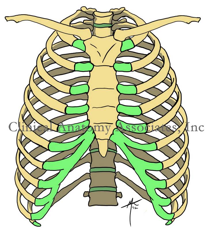
The thorax is the region of the trunk found between the neck superiorly and the abdomen inferiorly. It is characterized by a bony cage (thoracic cage). It is bound anteriorly by the sternum, laterally by twelve pairs of ribs and corresponding cartilages, and posteriorly by the thoracic spine, composed by twelve thoracic vertebrae.

Its superior boundary is known as the thoracic inlet or superior thoracic aperture, and it an oblique plane formed by the superior aspect of the sternum, the first rib, and the first thoracic vertebra. The inferior boundary is more difficult to describe, as there are two boundaries. The first is the inferior thoracic aperture or outlet formed by the lower end of the sternum, the cartilage of the costal margin and the most inferior ribs. The second one is the boundary between thorax and abdomen. formed by the respiratory diaphragm.

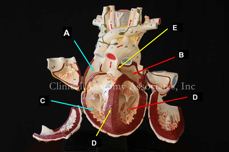
The thoracic cavity is divided into three separate regions; two laterally situated pleural regions, each containing a lung, and a centrally situated mediastinum.  The mediastinum contains the heart and pericardium, the trachea and bronchi, thoracic duct, thoracic esophagus, etc.

Image property of: CAA.Inc.Artist: D.M. Klein
Thorax, anterior view

Thorax - Anterior view. Click on the image for a larger version.

Back to MTD Main Page Subscribe to MTD

Anterior communicating artery

The anterior communicating artery is a single, unpaired artery found communicating both anterior cerebral arteries. The anterior communicating artery is one of the components of the arterial circle of Willis.

The anterior communicating artery is very short, usually between 2 to 3 mm in length, it is rarely absent, but it can be oblique or duplicated, or even triplicated. It is one of the sites for intracranial aneurysm. For more information on the anatomical variations of the anterior communicating artery click here.

For additional information:
"An Overview of Intracranial Aneurysms" Keedy, A Mcgill J Med. 2006 July; 9(2): 141–146 
Image modified from the original courtesy of Wikipedia.

Arterial circle of Willis  (Wikipedia.en.com
Back to MTD Main Page Subscribe to MTD

Ipsi-

The prefix [ipsi-] arises from the Latin [ipse] meaning "self" or "same". In medical terminology this prefix is used in the words "ipsilateral" and ipsilaterally" to denote an organ or structure in the "same side of the body" (left or right) as another structure that is being referred to.

A synonym for ipsilateral is [homolateral]; the antonym for the term is [contralateral].

Back to MTD Main Page Subscribe to MTD

Anterior cerebral artery

The anterior cerebral arteries are one of the two paired terminal branches of the internal carotid artery. Each anterior cerebral artery supplies arterial blood to the brain beyond the arterial circle of Willis. The vascular territory of the anterior cerebral artery supplies the medial surfaces of the frontal and parietal cerebral lobes as well as the medial border of the same lobes.

The anterior cerebral artery gives off the orbitofrontal artery and then divides into two callosal arteries, the pericallosal artery and the callosomarginal artery. From these arteries arise all the other branches that form the vascular territory of the anterior cerebral artery.

There are many anatomical variations of the anterior cerebral artery, as described here.

Anterior cerebral artery (ACA). a: artery; post: posterior; int: internal; sup: superior; inf:inferior. (Sobotta, 1945)
Clinical anatomy, pathology, and surgery of the brain and spinal cord are some of the lecture topics developed and delivered by Clinical Anatomy Associates, Inc.

Image modified from the original (in the public domain) by Sobotta (1945)

Back to MTD Main Page Subscribe to MTD