Medical Terminology Daily (MTD) is a blog sponsored by Clinical Anatomy Associates, Inc. as a service to the medical community. We post anatomical, medical or surgical terms, their meaning and usage, as well as biographical notes on anatomists, surgeons, and researchers through the ages. Be warned that some of the images used depict human anatomical specimens.

You are welcome to submit questions and suggestions using our "Contact Us" form. The information on this blog follows the terms on our "Privacy and Security Statement" and cannot be construed as medical guidance or instructions for treatment.


We have 77 guests online


A Moment in History

Jean-Louis Petit

Jean Louis Petit
(1674 – 1750)

French surgeon and anatomist, Jean Louis Petit was born in Paris in on March 13, 1674.  His family rented an apartment at his house to Alexis Littre (1658 – 1726), a French anatomist. Petit became an apprentice of Littre at seven years of age, helping him in the dissections for his lectures and at an early age became the assistant in charge of the anatomic amphitheater.

Because of Petit’s dedication to anatomy and medicine, in 1690 at the age of sixteen, became a disciple of a famous Paris surgeon, Castel.

In 1692, Petit entered the French army and performed surgery in two military campaigns. By 1693 he started delivering lectures and was accepted as a great surgeon, being invited to the most difficult operations.  In 1700 he was appointed Chief Surgeon of the Military School in Paris and in the same year he received the degree of Master of Surgery from the Faculty of Paris.

In 1715 he was made a member of the Royal Academy of Sciences and an honorary member of the Royal Society of London. He was appointed by the King as the first Director General of the Royal Academy of Surgery when it was founded in 1731.

Petit’s written works are of historical importance.  “Traite des Maladies des Os” ( A Treatise on Bone Diseases);  “Traite des Maladies Chirurgicales et des Operation” (A Treatise on Surgical Diseases and their Operations” This last book was published posthumously in 1774. He also published a monograph on hemorrhage, another on lachrymal fistula, and others.

He was one of the first to perform choIecystotomy and mastoidotomy. His original tourniquet design for amputations saved many in the battlefield and the design of the same surgical instrument today has not changed much since its invention by him.

His name is remembered in the lumbar triangle, also called the "triangle of Petit", and the abdominal hernia that can ensue through that area of weakness, the lumbar hernia or "Petit's hernia".

Sources:
1. “Jean Louis Petit – A Sketch of his Life, Character, and Writings” Hayne, AP San Fran Western Lancet 1875 4: 446-454
2. “Oeuvres compl?tes de Jean-Louis Petit” 1837 Imprimerie de F. Chapoulaud
3. Extraits de l'eloge de Jean-Louis Petit Ius dans Ia seance publique de I' Academie royale de chirurgie du 26 mai 1750” Louis A. Chirurgie 2001: 126 : 475- 81


 "Clinical Anatomy Associates, Inc., and the contributors of "Medical Terminology Daily" wish to thank all individuals who donate their bodies and tissues for the advancement of education and research”.

Click here for more information


abebooks banner

bookplateink.com


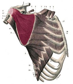
Subscapular muscle (subscapularis)

The subscapular muscle or subscapularis is a large triangular muscle which is found on the anterior aspect of the scapula, in close relation to the posterolateral aspect of the thorax. It is covered by a well-defined fascia layer, the subscapularis fascia. It is one of the muscles that forms the rotator cuff.

It originates from the internal aspect of the medial border of the scapula, in close proximity to the insertion of the serratus anterior (magnus), and the internal aspect of the inferolateral border of the scapula, where it is separated from the teres major muscle by a thick aponeurosis. It also takes origin directly from the subscapular fossa, where some of the muscular fibers attach directly to the bone.

The muscle inserts by way of a tendon in the lesser tubercle of the humerus and the anterior aspect of the glenohumeral joint capsule. The tendon of the muscle is separated from the neck of the scapula by a large bursa (the infratendinous bursa of the subscapularis) which communicates with the cavity of the glenohumeral joint through an aperture in the capsule.

It receives innervation by two subscapular nerves, both branches of the brachial plexus.

Subscapularis muscle - Image modified from the original by Henry VanDyke Carter, MD. Public domain
Subscapularis muscle.
Click on the image for a larger depiction 
The superior suprascapular nerve arises from the ventral rami of C5 and C6 nerve fibers. It branches from the posterior cord of the brachial plexus and supplies the superior aspect of the muscle. The inferior subscapular nerve arises from the ventral rami of C5 and C6 nerve fibers. It branches from the posterior cord of the brachial plexus and supplies the superior aspect of the muscle. Although these nerves have the same origin from the cervical spine, their origin from the posterior cord of the brachial plexus is different.

This muscle rotates the head of the humerus medially. When the upper extremity is raised, it draws the humerus anteroinferiorly.  As part of the shoulder’s rotator cuff it helps prevent subluxation of the glenohumeral joint by keeping the head of the humerus in situ.

The subscapularis is one of the 17 muscles that attach to the scapula.

Note: The image shown in this article is from “Gray’s Anatomy” (1918) which is in the public domain

Sources:
1. “Gray’s Anatomy” Henry Gray, 1918
2. "Tratado de Anatomia Humana" Testut et Latarjet 8th Ed. 1931 Salvat Editores, Spain
3. "Gray's Anatomy" 38th British Ed. Churchill Livingstone 1995
4. “An Illustrated Atlas of the Skeletal Muscles” Bowden, B. 4th Ed. Morton Publishing. 2015

Image modified from the original by Henry VanDyke Carter, MD. Public domain

MTD Main Page Subscribe to MTD

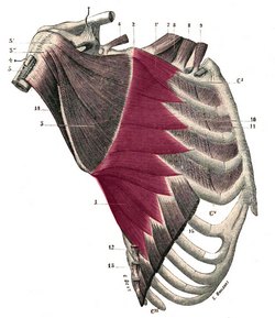
Serratus anterior (magnus)

The serratus anterior or serratus magnus is a is a wide, thin muscle sheet situated on the posterolateral aspect of the thorax and extends between the ribs and the scapula. It is formed by well-defined separate muscular digitations that originate in the external surface and superior aspect of the first superior eight (or nine) ribs. These originating fibers also arise from the fasciae covering the intercostal muscles. This is especially true for the first or most superior digitation which arises from the first and second rib and the intervening external intercostal fascia.

These digitations cover the lateral aspect of the thorax, pass deep to the scapula and converge to insert on the deep aspect of the medial border of the scapula. Some of its fibers may even hug the medial border of the scapula and insert on its anterior aspect. The first digitation is inserted into a triangular area on the ventral surface of the medial scapular angle. The next two digitations spread out to form a triangular sheet, the base of which is directed posteriorly and is inserted into nearly the whole length of the ventral surface of the vertebral border. The lower five or six digitations converge to form a fan-shaped mass, the apex of which inserts into a triangular impression on the ventral surface of the inferior scapular angle. The lower four slips of the serratus anterior interdigitate with the superior five muscular slips of the external oblique muscle.

Serratus magnus muscle - Image modified from the original by Henry VanDyke Carter, MD. Public domain
Serratus magnus muscle.
Click on the image for a larger depiction 
This muscle receives its nerve supply from the long thoracic nerve, (ventral rami of C5-C7), arising from the roots of C5, C6, and C7 (sometimes absent) of the brachial plexus

The word “serratus” derivates from the Latin word [serro] meaning “saw”. Serratus means “serrated” referring to the multiple tooth-like anterior digitations of the muscle. The plural form for "serratus" is " serrati". The Latin term “magnus” means “great”, “large”, or “mighty”. It points to the fact that this is the largest of three muscles that carry the same name “serratus”. The other two are the serratus posterior superior and the serratus posterior inferior.

The serratus anterior is one of the 17 muscles that attach to the scapula.

Note: The image shown in this article is from “Gray’s Anatomy” by Henry Gray (1918) which is in the public domain. It depicts the serratus anterior in situ and shows the scapula retracted posteriorly.  The scapula is covered on its internal aspect by the subscapularis muscle (number 3 in the image). A better image can be found in “An Illustrated Atlas of the Skeletal Muscles” by Bowden (2015) which we cannot publish for copyright reasons.

Sources:
1. “Gray’s Anatomy” Henry Gray, 1918
2. "Tratado de Anatomia Humana" Testut et Latarjet 8th Ed. 1931 Salvat Editores, Spain
3. "Gray's Anatomy" 38th British Ed. Churchill Livingstone 1995
4. “An Illustrated Atlas of the Skeletal Muscles” Bowden, B. 4th Ed. Morton Publishing. 2015

Image modified from the original by Henry VanDyke Carter, MD. Public domain

Back to MTD Main Page Subscribe to MTD

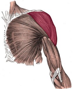
Deltoid muscle

The deltoid or deltoideus (Latin) is a large, thick, triangular muscle, which covers the glenohumeral joint anteriorly, superiorly, and posteriorly.  It can be described as having three components or segments, anterior, middle, and posterior. The anterior portion originates from the lateral third of the clavicle, on the clavicle’s superior border. The middle portion originates from the lateral border of the scapula’s acromion, and the posterior portion originates from the spine of the scapula. All three portions insert laterally by means of a thick tendon on the deltoid tuberosity of the humerus. At its insertion the muscle gives off an thick connective tissue expansion to the deep fascia of the arm.

The three portions of the deltoid muscle are usually well defined. As an anatomical variation, the clavicular or the acromial portion of the muscle may be absent.

This muscle is innervated by the axillary nerve (C5, C6), a branch of the brachial plexus, and supplied by the posterior circumflex humeral artery and the deltoid branch of the thoracoacromial artery.

The word deltoid is derivates from the Greek word [δελτοειδής] which is itself formed by the terms [δέλτα] (délta), referring to the triangular shape of the letter delta (uppercase Δ, lowercase δ) and [-οειδής] (-oeidís), a the Greek suffix meaning “similar to”. Delt-oid then would mean “similar to a Δ (delta)".

The deltoid is one of the 17 muscles that attach to the scapula.

Deltoid muscle - Image modified from the original by Henry VanDyke Carter, MD. Public domain
Deltoid muscle.
Click on the image for a larger depiction 
Sources:
1. “Gray’s Anatomy” Henry Gray, 1918
2. "Tratado de Anatomia Humana" Testut et Latarjet 8th Ed. 1931 Salvat Editores, Spain
3. "Gray's Anatomy" 38th British Ed. Churchill Livingstone 1995
4. “An Illustrated Atlas of the Skeletal Muscles” Bowden, B. 4th Ed. Morton Publishing. 2015

Image modified from the original by Henry VanDyke Carter, MD. Public domain

MTD Main Page Subscribe to MTD

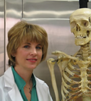
Dr. Elizabeth Murray Honored with American Academy of Forensic Sciences Award

We are proud to announce that our own contributor and associate Dr. Elizabeth Murray, Ph.D., has been awarded the Anthropology Section's 2018 "T. Dale Stewart Award" at the American Academy of Forensic Sciences (AAFS) 71st Annual Scientific Meeting in late February.

Dr. Murray's involvement with AAFS has included chairing the Academy-wide annual meeting as well as committee-level service in long-term planning, Board of Trustees, and the Student Academy.

Well-known and respected as a forensic anthropologist, Dr. Murray teaches at the University of Mount St. Joseph courses in anatomy and physiology, gross anatomy, and forensic science for the Department of Biology. Her most recent book, published in 2019, is "The Dozier School for Boys: Forensics, Survivors, and a Painful Past."

 dr murray
Our congratulations to her for yet another incredible achievement in her illustrious career. We are glad to count her as a friend and as a contributor to "Medical Terminology Daily" and Clinical Anatomy Associates, Inc.
Back to MTD Main Page Subscribe to MTD

Holly leaf sign

The Holly is a tree/shrub of the genus Ilex , with perhaps the most well know being Ilex aquifolium. The plant has shiny prickly evergreen leaves and bright red berries. Cut branches of Holly are widely used as a traditionally Christmas decoration especially in wreaths and Christmas cards as illustrations. “The Holly and the Ivy” is a popular traditional English Christmas carol.

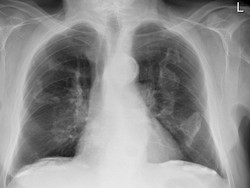
The [Holly leaf sign] refers to the appearance of calcified pleural plaques seen on chest radiographs. Pleural plaques are common in patients who have been exposed to asbestos, are asymptomatic and are most useful as a marker of asbestos exposure or asbestosis. They can be identified in 3-14% of dockyard workers and in 58% in insulation workers.

They are themselves not malignant, but patients with this plaques have a greater risk of mesothelioma and bronchogenic cancer than the general population and patients with exposed to asbestos but not pleural plaques.

Holly leaf sign - Case radiograph courtesy of Dr Çağlayan Çakır, Radiopaedia.org. From the case rID: 22986Holly leaf sign.
Click on the image for a larger depiction

The plaques arise in the parietal pleura and have predilection for the diaphragmatic dome and the undersurface of the lower posterolateral ribs. Rarely involve the visceral pleura but occasionally they are found in the fissures of the lungs.

On plain radiographic plaques appear as a geographic, usually calcified, opacities with irregular but well-defined edges. The irregular thickened nodular edges of the pleural plaques are likened to appearance of a Holly leaf, which has sharp spines along its margin.

Sources:
1. Jane R, Gulati A., Dwivedi R., Avula S., Curtis J., Abernethy L. (2013) We wish you a Merry X-Ray-mas: Christmas signs in radiology. BMJ 347:f7020 doi: 10.1136/bmj.f7020
2. Walker C., Takasugi J., Chung J., Reddy, G., Done S., Pipavath S., Schmidt R., Godwin J. (2012). Tumor-like Conditions of the Pleura. Radiographics 32:971–985.
3. Case radiograph courtesy of Dr Çağlayan Çakır, Radiopaedia.org. From the case rID: 22986

Figure below. Ilex aquifolium. Courtesy of A.Prof Frank Gaillard, Radiopaedia.org. From the case rID: 12398

Holly leaf sign - Ilex aquifolium. Courtesy of A.Prof Frank Gaillard, Radiopaedia.org. From the case rID: 12398
Holly leaf sign.
Click on the image for a larger depiction

Article submitted by: Prof. Claudio R. Molina, MsC..

Back to MTD Main Page Subscribe to MTD

Prostatic utricle

[UPDATED] The [prostatic utricle], also known as "utriculus prostaticus" or "utriculus" is a small 6 mm small dead-end channel found in the male prostatic urethra.

The word [utriculus] is Latin and means "little sac"..

What is interesting about this structure is that it is the embryological remnant in the male of the Müllerian ducts that form the vagina and the uterus in the female. In fact, in some texts the prostatic utricle is referred to as "uterus masculinus". Some researchers differ and point to the fact that this structure may not be a Müllerian duct derivate.

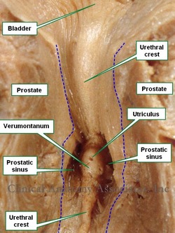
The prostatic utricle is found inside the prostate, forming part of the posterior wall of the prostatic urethra. It is in the upper part of a small mound which is part of the prostatic crest. This mound is called the [colliculus seminalis] or  [verumontanum], which is Latin and translates as the "mountain or mound of truth". On the verumontanum are the two slit-like openings of the ejaculatory ducts. Lateral to the verumontanum are the prostatic sinuses, depressions where the prostatic ducts are found.

Sources:
1. "The prostatic utricle is not a M?llerian duct remnant: immunohistochemical evidence for a distinct urogenital sinus origin" Shapiro E, Huang H, McFadden DE, et al. (2004) J Urol 172; 1753–1756
2. "Gray's Anatomy"38th British Ed. Churchill Livingstone 1995
3. "Tratado de Anatomia Humana" Testut et Latarjet 8 Ed. 1931 Salvat Editores, Spain

Anterior view of section of the prostate. The blue dotted line shows the edges of the prostatic urethra
    Anterior view of a section of the prostate gland. The blue dotted line shows the edges of the prostatic urethra.

Back to MTD Main Page Subscribe to MTD