|
The temporal fascia (Lat:Fascia Temporalis) is thick and strong muscular (deep) fascia that covers the external surface of the temporal muscle.
It originates on a curved line formed by the posterosuperior part of the zygomatic bone, the temporal line of the frontal bone, the upper temporal line of the temporal bone and the area between both upper and lower temporal lines. It is divided in two laminae: superficial and deep which have insertion on the zygomatic arch. The deep portion provides insertion to the temporal muscle (1,2). The superficial layer is part of the epicraneal aponeurosis (3).
The two layers of the temporal fasica have separate arterial and venous blodd supply.
Article written by: Maria F. Cortés, DDS, MSc.
Images from:
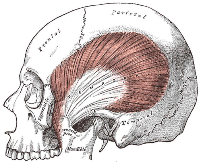
Fig 1. Public domain, by Henry Vandyke Carter, MD - Gray's Anatomy, 1918
|
Click on the image for a larger depiction
|
Sources:
1. “Anatomía humana” V.2. Latarjet- Ruiz Liard, 4ª ed. 6ª reimp. 2008 Médica Panamericana, Buenos Aires, Argentina.
2. “Anatomía humana: descriptiva, topográfica y funcional. Tomo 1. Cabeza y Cuello, Rouviere H – Delmas A, 11° ed. 2005 MASSON, S.A., Barcelona, Spain.
3. "Anatomy of the temporalis fascia" Wormald PJ, Alun-Jones T. J Laryngol Otol. 1991 Jul;105(7):522-4. |