Medical Terminology Daily (MTD) is a blog sponsored by Clinical Anatomy Associates, Inc. as a service to the medical community. We post anatomical, medical or surgical terms, their meaning and usage, as well as biographical notes on anatomists, surgeons, and researchers through the ages. Be warned that some of the images used depict human anatomical specimens.

You are welcome to submit questions and suggestions using our "Contact Us" form. The information on this blog follows the terms on our "Privacy and Security Statement" and cannot be construed as medical guidance or instructions for treatment.


We have 149 guests online


A Moment in History

Jean-Louis Petit

Jean Louis Petit
(1674 – 1750)

French surgeon and anatomist, Jean Louis Petit was born in Paris in on March 13, 1674.  His family rented an apartment at his house to Alexis Littre (1658 – 1726), a French anatomist. Petit became an apprentice of Littre at seven years of age, helping him in the dissections for his lectures and at an early age became the assistant in charge of the anatomic amphitheater.

Because of Petit’s dedication to anatomy and medicine, in 1690 at the age of sixteen, became a disciple of a famous Paris surgeon, Castel.

In 1692, Petit entered the French army and performed surgery in two military campaigns. By 1693 he started delivering lectures and was accepted as a great surgeon, being invited to the most difficult operations.  In 1700 he was appointed Chief Surgeon of the Military School in Paris and in the same year he received the degree of Master of Surgery from the Faculty of Paris.

In 1715 he was made a member of the Royal Academy of Sciences and an honorary member of the Royal Society of London. He was appointed by the King as the first Director General of the Royal Academy of Surgery when it was founded in 1731.

Petit’s written works are of historical importance.  “Traite des Maladies des Os” ( A Treatise on Bone Diseases);  “Traite des Maladies Chirurgicales et des Operation” (A Treatise on Surgical Diseases and their Operations” This last book was published posthumously in 1774. He also published a monograph on hemorrhage, another on lachrymal fistula, and others.

He was one of the first to perform choIecystotomy and mastoidotomy. His original tourniquet design for amputations saved many in the battlefield and the design of the same surgical instrument today has not changed much since its invention by him.

His name is remembered in the lumbar triangle, also called the "triangle of Petit", and the abdominal hernia that can ensue through that area of weakness, the lumbar hernia or "Petit's hernia".

Sources:
1. “Jean Louis Petit – A Sketch of his Life, Character, and Writings” Hayne, AP San Fran Western Lancet 1875 4: 446-454
2. “Oeuvres compl?tes de Jean-Louis Petit” 1837 Imprimerie de F. Chapoulaud
3. Extraits de l'eloge de Jean-Louis Petit Ius dans Ia seance publique de I' Academie royale de chirurgie du 26 mai 1750” Louis A. Chirurgie 2001: 126 : 475- 81


 "Clinical Anatomy Associates, Inc., and the contributors of "Medical Terminology Daily" wish to thank all individuals who donate their bodies and tissues for the advancement of education and research”.

Click here for more information


abebooks banner

bookplateink.com


Dextrocardia

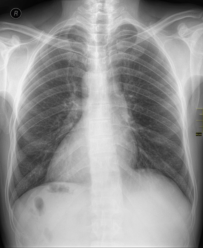
Chest X-Ray - dextrocardia

Normally, the heart is a midline structure found just posterior to the sternum where 35-40% of the heart mass in the right side of the thorax (chest), and the rest (60-65%) is in the left side of the thorax. In this normal condition the apex of the heart faces slightly inferior and to the left. In fact, there are many books and websites that state (wrongly) that the heart is normally in the “left side of the chest”.

If the above mentioned situation is reversed, we are in the presence of dextrocardia, that is, the heart is still in the midline, but most of the mass of the heart is in the right side of the thorax, and the apex points inferiorly and to the right.

The word dextrocardia is a derivate of the Latin [dexter], meaning “right”, and the Greek term [kardia], meaning “heart”. The word dextrocardia literally means “right-sided heart”.

Dextrocardia is a congenital condition, can be completely asymptomatic and present as an isolated condition. It can also be part of a complex genetic condition called “situs inversus” where the whole body is a mirror image of itself and all organs, including the heart are mirrored. A complete situs inversus is rare, but when present it usually does not cause problems.

The problems start when only part of the body and organs are reversed and others are not, causing an incredible number of potential anatomical variations and associated problems.

The prevalence of dextrocardia is about 1 in 12,00 pregnancies. The reported incidence is about 0.22%. Depending on the situation, dextrocardia can present with additional cardiac congenital disorders.

Sources

1. “Dextrocardia: an incidental finding” Yusuf SW, Durand JB, Lenihan DJ, Swafford J. Tex Heart Inst J 2009;36(4):358-9.
2. Garg N, Agarwal BL, Modi N, Radhakrishnan S, Sinha N. Dextrocardia: an analysis of cardiac structures in 125 patients. Int J Cardiol 2003;88(2–3):143–56
3. Bernasconi A, Azancot A, Simpson JM, Jones A, Sharland GK. Fetal dextrocardia: diagnosis and outcome in two tertiary centres. Heart 2005;91(12):1590–4.