Medical Terminology Daily (MTD) is a blog sponsored by Clinical Anatomy Associates, Inc. as a service to the medical community. We post anatomical, medical or surgical terms, their meaning and usage, as well as biographical notes on anatomists, surgeons, and researchers through the ages. Be warned that some of the images used depict human anatomical specimens.

You are welcome to submit questions and suggestions using our "Contact Us" form. The information on this blog follows the terms on our "Privacy and Security Statement" and cannot be construed as medical guidance or instructions for treatment.


We have 217 guests online


A Moment in History

Jean-Louis Petit

Jean Louis Petit
(1674 – 1750)

French surgeon and anatomist, Jean Louis Petit was born in Paris in on March 13, 1674.  His family rented an apartment at his house to Alexis Littre (1658 – 1726), a French anatomist. Petit became an apprentice of Littre at seven years of age, helping him in the dissections for his lectures and at an early age became the assistant in charge of the anatomic amphitheater.

Because of Petit’s dedication to anatomy and medicine, in 1690 at the age of sixteen, became a disciple of a famous Paris surgeon, Castel.

In 1692, Petit entered the French army and performed surgery in two military campaigns. By 1693 he started delivering lectures and was accepted as a great surgeon, being invited to the most difficult operations.  In 1700 he was appointed Chief Surgeon of the Military School in Paris and in the same year he received the degree of Master of Surgery from the Faculty of Paris.

In 1715 he was made a member of the Royal Academy of Sciences and an honorary member of the Royal Society of London. He was appointed by the King as the first Director General of the Royal Academy of Surgery when it was founded in 1731.

Petit’s written works are of historical importance.  “Traite des Maladies des Os” ( A Treatise on Bone Diseases);  “Traite des Maladies Chirurgicales et des Operation” (A Treatise on Surgical Diseases and their Operations” This last book was published posthumously in 1774. He also published a monograph on hemorrhage, another on lachrymal fistula, and others.

He was one of the first to perform choIecystotomy and mastoidotomy. His original tourniquet design for amputations saved many in the battlefield and the design of the same surgical instrument today has not changed much since its invention by him.

His name is remembered in the lumbar triangle, also called the "triangle of Petit", and the abdominal hernia that can ensue through that area of weakness, the lumbar hernia or "Petit's hernia".

Sources:
1. “Jean Louis Petit – A Sketch of his Life, Character, and Writings” Hayne, AP San Fran Western Lancet 1875 4: 446-454
2. “Oeuvres compl?tes de Jean-Louis Petit” 1837 Imprimerie de F. Chapoulaud
3. Extraits de l'eloge de Jean-Louis Petit Ius dans Ia seance publique de I' Academie royale de chirurgie du 26 mai 1750” Louis A. Chirurgie 2001: 126 : 475- 81


 "Clinical Anatomy Associates, Inc., and the contributors of "Medical Terminology Daily" wish to thank all individuals who donate their bodies and tissues for the advancement of education and research”.

Click here for more information


abebooks banner

bookplateink.com


The lost influence of Andreas Vesalius on eponymic anatomy

Andreas Vesalius Bruxellensis (1515-1564) is considered to be the father of modern anatomy, remembered because an illustrious life and by his book “De Humani Corporis Fabrica, Libri Septem” published first in 1543. Scores of books, translations, articles, and analyzes of his work have been published in the over 500 years since his birth.

With such an incredible pedigree we would expect his name to be attached to more than a few anatomical structures, many of which were first described in his opus magnus, the “Fabrica”, I wonder why this is not so. It is true that modern anatomy is trying to move from eponyms to more descriptive anatomical terms. Still, there are many that will not go away, as is the famous ligament of Treitz, or the sphincter or Oddi.

Today there is only one recorded eponym for Andreas Vesalius, the os vesalianum, a rare accessory bone in the foot located proximally to the base of the fifth metatarsal. It is usually asymptomatic, but in rare cases it can cause pain. It is formed by the failed fusion of the secondary ossification center of the fifth metatarsal.

Reviewing history, I was able to find other references to Andreas Vesalius eponyms or potential eponyms, now in disuse or misnamed:

Andreas Vesalius Bruxellensis
Suspensory ligaments of the mammary gland. Although first described by Vesalius in the Fabrica, these structures are named after Sir Astley Paston Cooper (1768 -1841), almost 300 years after being described by the great anatomist, who called them a “fleshy membrane” that stretched between the pectoral fascia and the skin.

- The vermiform appendix. Although called by many just “the appendix”, this structure is mentioned, but not named, by Jacobo Berengario da Carpi in 1524. It was Andreas Vesalius who first described it as an appendix, suggested it looked like a worm (Lat: vermis) calling it the “vermiform appendix”.

- The ligamentum suspensorium Vesalii or crural arch. First described by Giovanni Baptista Morgagni (1682-1771), it was named in honor of Vesalius by Dr. Laurentii (Lorenz) Heisters in his “Compendium Anatomicum” published in 1756. Other authors point to Gabrielle Fallopius as the first to describe this structure in 1561, although he did publish later than Vesalius (1543). Although named after Vesalius, it was later named after Francois Poupart who described it in 1695. You probably know this structure as the inguinal ligament.

-The ligamentum teres femoris. The round ligament of the femur was also first described by Vesalius in 1543.

NOTE: If you have other structures that have been named after Vesalius, please let me know by clicking here.

Sources:
1. “A Rare Cause of Foot Pain with Golf Swing. Symptomatic Os Vesalianum Pedis—A Case Report” Petrera, M et al. Sports Health. 2013 Jul; 5(4): 357–359.
2. “Andreas Vesalius’ 500th Anniversary: First Description of the Mammary Suspensory Ligaments” Brinkman RJ, Hage, JJ. World J Surg (2016) 40:2144–2148
3. “Compendium Anatomicum” Heisters, L. 1756 (German)
4. “Anatomy: An Encyclopedic Reference to the Language of Anatomy and Neuroanatomy. It Provides the Fascinating Origin of Terms and Biographies of Anatomists/Physicians Who Originated Them” Bergman, RA, Afifi, AK 2016

MTD Main Page Subscribe to MTD

Eponym

UPDATED: From the Greek [ep(i)] meaning "outer, above, or upon", and the Greek suffix [o-nym] meaning "name". The word [eponym] refers to a person's name becoming attached to an anatomical location or surgical procedure. For centuries it has been the custom to honor or remember someone by attaching their name to a structure, location, procedure, or maneuver.

This has changed as anatomists tend now to give locations and structures descriptive terms. An example of this would be the "Ampulla of Vater" named after the German anatomist Abraham Vater (1684-1751) described today in anatomical texts as the "hepatopancreatic ampulla". The controversy on using eponyms or not goes on...

There are many eponymical terms in the medical arena; following are some of them, click on the links for additional information:

Hesselbach’s triangle: Named after Franz Kaspar Hesselbach (1759-1816) (see yellow insert in superior image)
Spigelian line (linea semilunaris): Site for an Spigelian hernia, named after Adrian Van Der Spigelius (1578-1625) (see blue arrow in inferior image)
Fallopian tube: Named after Gabrielle Fallopius (1523-1563)
Cooper's pectineal ligament. Named after Sir Astley Paston Cooper (1768-1841)
Hartmann's procedure: A two-stage colon resection and anastomosis. Named after Prof. Henri Hartmann (1860-1952), a French surgeon.
Heimlich's maneuver:  Named after Dr. Henry J. Heimlich (1920 - )
Ligament of Treitz: Named after Václav Treitz (1819 - 1872), a Czech pathologist.

If you want to see a listing of the eponyms in this website, click here.

Here is an article on "The lost influence of Andreas Vesalius on eponymic anatomy".

Here is an interesting article on eponyms by Ilana Yurkiewicz published on 11/15/2012 in Scientific American: "Modern medical terms are still named after Nazi doctors. Can we change it?". It is interesting and thougthful reading.

PERSONAL NOTE: Many anatomists today are actively trying to eliminate eponyms from anatomical, medical, and surgical books. For me, this eliminates the interest of learning about the people who either first described these structures or procedures, which is one of the objectives of this website. I wonder (and this is a tongue-in-cheek comment) if the reason for this desire to eliminate eponyms is because there are so many attached to anatomical structures that there is no place for their own names! When history has forgotten about the original eponyms maybe we will see new ones with the names of modern anatomists! I do not worry, my name is attached to the "Ligaments of Miranda". Dr. Miranda

Superior image property of: CAA.Inc.. Artist: M. Zuptich.
Inferior image property of:CAA.Inc.. Artist:D.M. Klein

Back to MTD Main Page Subscribe to MTD

Ascending aorta

Ascending aorta, anterior view
Ascending aorta

UPDATED: The ascending aorta is the first and most proximal portion of the aorta. About 5 cm. in length and 3 cm. in diameter at its origin, its proximal end begins at the superior aspect of the outflow tract of of the left ventricle, at the ventriculoaortic junction. 

The ascending aorta ends superiorly at an imaginary horizontal plane (blue dotted line) that passes through the sternal angle (of Louis), continuing distally with the aortic arch. This is an important anatomical landmark, as many surgeons use as the superior border of the ascending aorta an oblique plane that passes proximal to the brachiocephalic trunk (yellow dotted line). Although this landmark could be useful in surgery, it is not anatomically correct.

Since the sternal angle (of Louis) also indicates the superior border of the pericardial sac, it can be said that the ascending aorta is completely intrapericardial, and in surgery the pericardial sac should be the anatomical landmark used to separate the ascending aorta from the aortic arch.

From its point of origin at the ventriculoaortic junction, the aorta presents with a dilated region where the aortic valve is located. The aortic valve is one of the two  semilunar valves of the heart, and the dilation of this region is caused by the presence of the sinuses of Valsalva. This dilated bulbous segment is known as the aortic root.

The dilated, sinus portion, or aortic root segment of the ascending aorta continues superiorly with the tubular portion of the ascending aorta. The area of transition between these two components is marked by a sharp crease known as the sinotubular junction (STJ). The dilation of the aortic root is caused by the presence of the sinuses of Valsalva, named after Antonio Maria Valsalva (1666 - 1723).

The tubular portion of the ascending aorta ascends with an inclination anteriorly and to the right. The ascending aorta presents with a slight anterior bulge causing the transverse section of this aortic segment to be slightly oval.

Only two arteries arise from the ascending aorta, both usually at the aortic root segment, just inferior to the STJ. These are the right coronary artery and the left coronary artery. There are anatomical variations where only one, or up to five different coronary arteries have been described.

Image property of:CAA.Inc.Artist:Dr. E. Miranda


Kernicterus

Kernicterus is a disorder where excess bilirubin accumulates in the deep neural tissues of the brain and can cause brain damage in the newborn.

It is characterized by jaundice and a limpness of the newborn, devoid of energy. Can present with seizures, convulsions, and muscle spasms.

This condition is treatable and requires awareness from the parents if yellowness of the skin (jaundice) is detected along with the above-mentioned signs in the early days post-partum. There are other signs not mentioned in this article

The word [kernicterus] comes from the German word [kern], meaning “nucleus” or “core”. In this particular word the term kern refers to the fact that one of the most importantly affected brain structures in kernicterus are the basal ganglia of the brain (also known as the "central nuclei", found at the "core" of the brain. It also includes the word [icterus] from the Greek word [ικτερός] pronounced (ikterós). The word [icterus] in Greek was originally used to denote a yellow bird, and is now used to denote the yellow color of jaundice.

 Newborn with kernicterus jaundice
Click on the image for a larger version. 

We would like to thank diseasepictures.com for the image in this article. For additional information on neonatal jaundice, click here.

Sources:
1. Clayman, L. "The AMA Encyclopedia of Medicine" 1989. Random House, NY
2. “The Origin of Medical Terms” Skinner HA 1970 Hafner Publishing Co.

Thanks to Jackie Miranda-Klein for her contribution suggesting this word.

MTD Main Page Subscribe to MTD

Parenchyma

The term [parenchyma] is a Greek term (παράένχέω). Its origin and meaning have little relation to the medical use of the term. The word means "that what is poured" or to "pour in". The actual definition of the term is "the proper mass of a solid organ". If someone refers to the "liver parenchyma", they are referring to the hepatic tissue, so it is with any other solid organ.

The etymology of the word is obscure and reflects ancient physiological theories and history. Vesalius mentions that the word was introduced by Erasistratus circa 300BC. He thought that the blood was "poured" into the organ and then this poured fluid would congeal to form the organ's proper mass. With time this concept was abandoned, but the word persisted to its modern meaning.

Interesting, there are many which accentuate the word wrongly. The accent or stress should be on the letter "e" and not on the letter 'y", so it should be pronounced "parénchyma"

Back to MTD Main Page Subscribe to MTD

Layers of the GI tract

Layers of the gastrointestinal tract
Layers of the GI tract

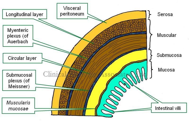
The gastrointestinal (GI) tract is formed, with a few exceptions, by four concentric layers of tissue. These are, from deep to superficial, the mucosa, submucosa, muscular (or muscularis) and the serosa layers. This is the simplified version. The fact is that there are more sublayers.

The mucosa layer is characterized by the presence of intestinal villi, which in the stomach and small intestine contribute to absorption of the digested food. The mucosa has a thin layer of connective called the "lamina propia" and external to it a thin layer of smooth muscle, the muscularis mucosae.

The submucosa layer is formed by irregular connective tissue and contains on its most external region a plexus of nerves and neurons, the "submucosal plexus of Meissner", which provides parasympathetic innervation to glands and the muscularis mucosae.

The muscular layer, also known as the "muscularis" is composed of two sublayers of smooth muscle. The deep layer contains circular fibers and is known either as the "circular muscle layer" or the "muscularis interna", the superficial layer contains longitudinal smooth muscle fibers and is known as the "longitudinal muscle layer" or the muscularis externa. Between both muscle layers lies the "myenteric plexus of Auerbach", a layer of sympathetic and parasympathetic nerves and neurons that provides nerve supply to the muscular layer. The combined action of this plexus on the muscular layer is responsible for peristalsis.

The serosa layer is the outer or external layer and is formed by a layer of peritoneum. As such, this layer can also be called "visceral peritoneum".

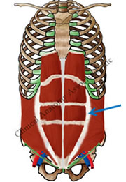
There are variations from GI organ to GI organ in the arrangement, content, glands, thickness of the layers, etc. The most important differences can be found in the thoracic esophagus and most of the rectum which are devoid of a serosa layer, and in the stomach, where there is a third muscular layer, deep to the circular layer, called the "oblique layer" that contributes fibers to the lower esophageal sphincter found at the esophagogastric junction.

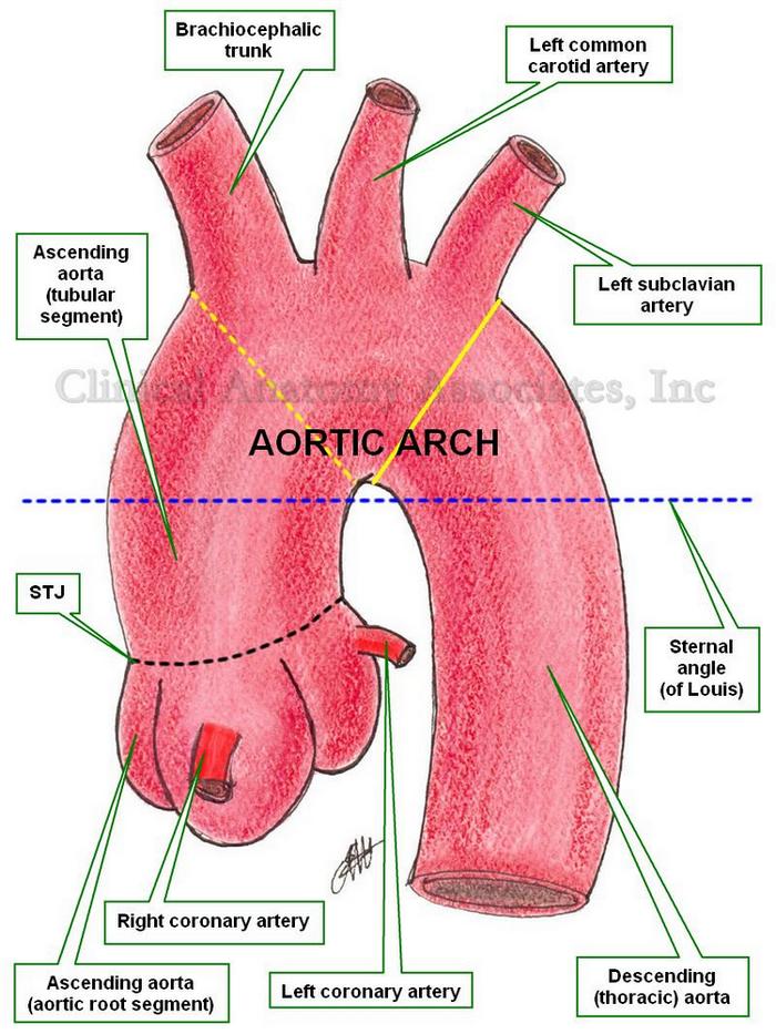
An important point to make is the presence of two interconnected ganglionated plexuses that are represented in the GI tract by the submucosal plexus of Meissner and the myenteric plexus of Auerbach which form the GI intrinsic autonomic nervous component . These two plexuses extend from the esophagus to the rectum and allow for the GI tract to operate almost independently from the extrinsic autonomic nervous system which moderates their activity. Ganglionated plexuses are present in organs that have rhythmic activity, such as peristalsis. Ganglionated plexuses are also present in the heart.

Sources:
1. "The bowel and beyond: the enteric nervous system in neurological disorders" Rao, M & Gershon, M. Nat Rev Gastroenterol Hepatol. 2016 Sep; 13(9): 517–528. 
2. "Advances in Enteric Neurobiology: The “Brain” in the Gut in Health and Disease" Kulkami, S et al. Journal of Neuroscience 31 October 2018, 38 (44) 9346-9354
3. "The Brain-Gut Connection" John Hopklins Health
4. "Think Twice: How the Gut's "Second Brain" Influences Mood and Well-Being" Hadhazy, B. Scientific American February 2010

Images property of:CAA.Inc.Artist:Dr. E. Miranda