|
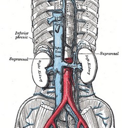
The inferior vena cava (IVC) in one of the great vessels. It brings deoxygenated blood from the lower extremities, pelvis, and areas of the abdomen to the right atrium of the heart. As a side note, the blood returning from the digestive system does not usually enter the IVC. It has it its own venous subsystem converging into the liver by way of the portal vein. The IVC is formed by the confluence of the right and left common iliac veins. This lower end of the inferior vena cava is found anterior to the L4-L5 intervertebral disc. The IVC covers the superior aspect of the body of L5. The IVC ascends to the right of the abdominal aorta and anterolateral to the vertebral bodies. It receives several branches as it passes superiorly: • Common iliac veins |
|
| As the IVC passes posterior to the liver, it is hugged by the mass of the posterior aspect of the liver, it will receive the hepatics veins, and pass through the IVC hiatus of the respiratory diaphragm, entering immediately into the right atrium of the heart. At this point the IVC will present an incomplete venous valve known as the Eustachian valve, named after Bartolomeo Eustachius (c1500 - 1574).
Sources: |
|
| Back to MTD Main Page | Subscribe to MTD |
