Medical Terminology Daily (MTD) is a blog sponsored by Clinical Anatomy Associates, Inc. as a service to the medical community. We post anatomical, medical or surgical terms, their meaning and usage, as well as biographical notes on anatomists, surgeons, and researchers through the ages. Be warned that some of the images used depict human anatomical specimens.

You are welcome to submit questions and suggestions using our "Contact Us" form. The information on this blog follows the terms on our "Privacy and Security Statement" and cannot be construed as medical guidance or instructions for treatment.


We have 152 guests online


A Moment in History

Jean-Louis Petit

Jean Louis Petit
(1674 – 1750)

French surgeon and anatomist, Jean Louis Petit was born in Paris in on March 13, 1674.  His family rented an apartment at his house to Alexis Littre (1658 – 1726), a French anatomist. Petit became an apprentice of Littre at seven years of age, helping him in the dissections for his lectures and at an early age became the assistant in charge of the anatomic amphitheater.

Because of Petit’s dedication to anatomy and medicine, in 1690 at the age of sixteen, became a disciple of a famous Paris surgeon, Castel.

In 1692, Petit entered the French army and performed surgery in two military campaigns. By 1693 he started delivering lectures and was accepted as a great surgeon, being invited to the most difficult operations.  In 1700 he was appointed Chief Surgeon of the Military School in Paris and in the same year he received the degree of Master of Surgery from the Faculty of Paris.

In 1715 he was made a member of the Royal Academy of Sciences and an honorary member of the Royal Society of London. He was appointed by the King as the first Director General of the Royal Academy of Surgery when it was founded in 1731.

Petit’s written works are of historical importance.  “Traite des Maladies des Os” ( A Treatise on Bone Diseases);  “Traite des Maladies Chirurgicales et des Operation” (A Treatise on Surgical Diseases and their Operations” This last book was published posthumously in 1774. He also published a monograph on hemorrhage, another on lachrymal fistula, and others.

He was one of the first to perform choIecystotomy and mastoidotomy. His original tourniquet design for amputations saved many in the battlefield and the design of the same surgical instrument today has not changed much since its invention by him.

His name is remembered in the lumbar triangle, also called the "triangle of Petit", and the abdominal hernia that can ensue through that area of weakness, the lumbar hernia or "Petit's hernia".

Sources:
1. “Jean Louis Petit – A Sketch of his Life, Character, and Writings” Hayne, AP San Fran Western Lancet 1875 4: 446-454
2. “Oeuvres compl?tes de Jean-Louis Petit” 1837 Imprimerie de F. Chapoulaud
3. Extraits de l'eloge de Jean-Louis Petit Ius dans Ia seance publique de I' Academie royale de chirurgie du 26 mai 1750” Louis A. Chirurgie 2001: 126 : 475- 81


 "Clinical Anatomy Associates, Inc., and the contributors of "Medical Terminology Daily" wish to thank all individuals who donate their bodies and tissues for the advancement of education and research”.

Click here for more information


abebooks banner

bookplateink.com


Lung

A [lung] is one of the two main respiratory organs. It is an organ filled with minute air sacs or alveoli, giving its parenchyma the look and feel of a spongy tissue. Its function is the reoxygenation of blood.

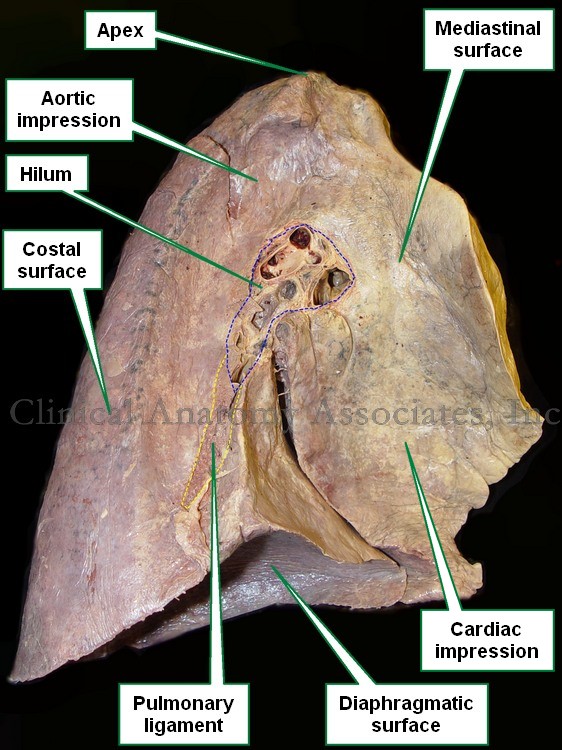
Each lung is located on each side of the thoracic cavity, each one surrounded by a serosa membrane called "pleura". The pleura forms a sac that surrounds the lung and also lines the thoracic wall. The pleura that lines the thoracic wall is called "parietal pleura", while the pleura that lines the lung is called the "visceral pleura". An extension of the visceral pleura inferior to the pulmonary hilum forms the pulmonary ligament.

A lung has three surfaces and an apex. The inferior surface, base, or diaphragmatic surface; the medial surface or mediastinal surface, as if forms the lateral wall of the mediastinum; and the lateral or costal surface.

The lungs are also divided into lobes. The right lung has three lobes: superior, middle, and inferior while the left lung only has two lobes: superior and inferior. Because the heart has a left-sided tilt the left lung is slightly smaller than the right lung.

There are four structures that enter or leave the lung and they are found at the pulmonary hilum in the medial surface of the lung. These structures are: the main or primary bronchus, the pulmonary artery, and two pulmonary veins.

Medial surface of a left lung
Image property of: CAA.Inc.Photographer:David M. Klein
After entering the lung through the hilum each bronchus will divide into smaller and smaller bronchi eventually opening into microscopic air sacs called the alveoli. Each alveolus or alveolar sac is surrounded by capillaries. It is here where gas exchange takes place allowing for blood reoxygenation.

When examined, a lung will show the impressions of the organs that are in contact with it. Click on the accompanying image and you will be able to see the impressions left by the aorta and the heart

Back to MTD Main Page Subscribe to MTD