Sponsor   

Medical Terminology Daily (MTD) is a blog sponsored by Clinical Anatomy Associates, Inc. as a service to the medical community, medical students, and the medical industry. We post anatomical, medical or surgical terms, their meaning and usage, as well as biographical notes on anatomists, surgeons, and researchers through the ages. Be warned that some of the images used depict human anatomical specimens.

Click on the link below to subscribe to the MTD newsletter. If you think an article could be interesting to somebody else, feel free to forward the link of the article. Should you want to use the information on the article, please follow the CAA, Inc Privacy and Security Statement found at the bottom of this page. 

You are welcome to submit questions and suggestions using our "Contact Us" form. The information on this blog follows the terms on our "Privacy and Security Statement"  and cannot be construed as medical guidance or instructions for treatment. 


We have 51 guests online


A Moment in History

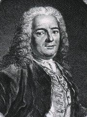
Self-portrait, Henry Vandyke Carter, MD (Public Domain)

Henry Vandyke Carter, MD
(1831 – 1897)

English physician, surgeon, medical artist, and a pioneer in leprosy and mycetoma studies.  HV Carter was born in Yorkshire in 1831. He was the son of Henry Barlow Carter, a well-known artist and it is possible that he honed his natural talents with his father. His mother picked his middle name after a famous painter, Anthony Van Dyck. This is probably why his name is sometimes shown as Henry Van Dyke Carter, although the most common presentation of his middle name is Vandyke.

Having problems to finance his medical studies, HV Carter trained as an apothecary and later as an anatomical demonstrator at St. George’s Hospital in London, where he met Henry Gray (1872-1861), who was at the time the anatomical lecturer. Having seen the quality of HV Carter’s drawings, Henry Gray teamed with him to produce one of the most popular and longer-lived anatomy books in history: “Gray’s Anatomy”, which was first published in late 1857.  The book itself, about which many papers have been written, was immediately accepted and praised because of the clarity of the text as well as the incredible drawings of Henry Vandyke Carter.

While working on the book’s drawings, HV Carter continued his studies and received his MD in 1856.

In spite of initially being offered a co-authorship of the book, Dr. Carter was relegated to the position of illustrator by Henry Gray and never saw the royalties that the book could have generated for him. For all his work and dedication, Dr. Carter only received a one-time payment of 150 pounds. Dr.  Carter never worked again with Gray, who died of smallpox only a few years later.

Frustrated, Dr. Carter took the exams for the India Medical Service.  In 1858 he joined as an Assistant Surgeon and later became a professor of anatomy and physiology. Even later he served as a Civil Surgeon. During his tenure with the India Medical Service he attained the ranks of Surgeon, Surgeon-Major, Surgeon-Lieutenant-Colonel, and Brigade-Surgeon.

Dr. Carter dedicated the rest of his life to the study of leprosy, and other ailments typical of India at that time. He held several important offices, including that of Dean of the Medical School of the University of Bombay. In 1890, after his retirement, he was appointed Honorary Physician to the Queen.

Dr. Henry Vandyke Carter died of tuberculosis in 1897.

Personal note: Had history been different, this famous book would have been called “Gray and Carter’s Anatomy” and Dr. Carter never gone to India. His legacy is still seen in the images of the thousands of copies of “Gray’s Anatomy” throughout the world and the many reproductions of his work available on the Internet. We are proud to use some of his images in this blog. The image accompanying this article is a self-portrait of Dr. Carter. Click on the image for a larger depiction. Dr. Miranda

Sources:
1. “Obituary: Henry Vandyke Carter” Br Med J (1897);1:1256-7
2. “The Anatomist: A True Story of ‘Gray’s Anatomy” Hayes W. (2007) USA: Ballantine
3. “A Glimpse of Our Past: Henry Gray’s Anatomy” Pearce, JMS. J Clin Anat (2009) 22:291–295
4. “Henry Gray and Henry Vandyke Carter: Creators of a famous textbook” Roberts S. J Med Biogr (2000) 8:206–212.
5. “Henry Vandyke Carter and his meritorious works in India” Tappa, DM et al. Indian J Dermatol Venereol Leprol (2011) 77:101-3


"Clinical Anatomy Associates, Inc., and the contributors of "Medical Terminology Daily" wish to thank all individuals who donate their bodies and tissues for the advancement of education and research”.

Click here for more information


Rare & Collectible Books at AbeBooks.com 

bookplateink.com


Trapezius

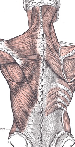
The [trapezius] is a bilateral muscle belonging to the superficial muscles of the back. On each side it is a flat, thin triangular muscle that spans the neck, shoulders and the superior and middle aspect of the back. When seen together these two triangular muscles form a diamond-shaped quadrangle from which its name derives. The word originates in the Greek [τραπεζι] meaning "a four-legged table" (four sides). This word later evolved into the New Latin [trapezium].

In the midline the trapezius muscle attaches to the inion (external occipital protuberance), the ligamentum nuch?, the spinous processes of the seventh cervical vertebra (vertebra prominens), and the spinous processes of all the thoracic vertebr?.

The trapezius’ muscle fibers have three orientations. From the midline the superior fibers course inferolaterally to attach to the posterior border of the lateral third of the clavicle. The middle fibers course laterally to attach to the medial margin of the acromion, and posterior border of the spine of the scapula. The inferior fibers course superolaterally to attach to the spine of the scapula by way of an aponeurosis.

Because of their attachments, the superior and inferior fibers of the trapezius act coordinatedly to rotate the scapula, while the middle fibers act to retract the scapula. The superior fibers also act to slightly elevate the scapula. The trapezius muscle is sometimes described as an accessory respiratory muscle.

The trapezius muscle receives muscular innervation by way of the spinal accessory nerve (11th Cranial Nerve) which courses on the deep aspect of the muscle along with the  superficial branch of the transverse cervical artery and vein. The muscle also receives sensory innervation by way of nerves arising from the ventral rami of the 3rd and 4th spinal nerves.

The trapezius is one of the 17 muscles that attach to the scapula.

Image courtesy of Bartleby.com 

Posterior view of the superficial and intermediate muscle layers of the back (bartleby.com)