Medical Terminology Daily (MTD) is a blog sponsored by Clinical Anatomy Associates, Inc. as a service to the medical community. We post anatomical, medical or surgical terms, their meaning and usage, as well as biographical notes on anatomists, surgeons, and researchers through the ages. Be warned that some of the images used depict human anatomical specimens.

You are welcome to submit questions and suggestions using our "Contact Us" form. The information on this blog follows the terms on our "Privacy and Security Statement" and cannot be construed as medical guidance or instructions for treatment.


We have 307 guests online


A Moment in History

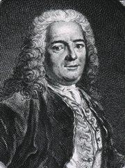
Jean-Louis Petit

Jean Louis Petit
(1674 – 1750)

French surgeon and anatomist, Jean Louis Petit was born in Paris in on March 13, 1674.  His family rented an apartment at his house to Alexis Littre (1658 – 1726), a French anatomist. Petit became an apprentice of Littre at seven years of age, helping him in the dissections for his lectures and at an early age became the assistant in charge of the anatomic amphitheater.

Because of Petit’s dedication to anatomy and medicine, in 1690 at the age of sixteen, became a disciple of a famous Paris surgeon, Castel.

In 1692, Petit entered the French army and performed surgery in two military campaigns. By 1693 he started delivering lectures and was accepted as a great surgeon, being invited to the most difficult operations.  In 1700 he was appointed Chief Surgeon of the Military School in Paris and in the same year he received the degree of Master of Surgery from the Faculty of Paris.

In 1715 he was made a member of the Royal Academy of Sciences and an honorary member of the Royal Society of London. He was appointed by the King as the first Director General of the Royal Academy of Surgery when it was founded in 1731.

Petit’s written works are of historical importance.  “Traite des Maladies des Os” ( A Treatise on Bone Diseases);  “Traite des Maladies Chirurgicales et des Operation” (A Treatise on Surgical Diseases and their Operations” This last book was published posthumously in 1774. He also published a monograph on hemorrhage, another on lachrymal fistula, and others.

He was one of the first to perform choIecystotomy and mastoidotomy. His original tourniquet design for amputations saved many in the battlefield and the design of the same surgical instrument today has not changed much since its invention by him.

His name is remembered in the lumbar triangle, also called the "triangle of Petit", and the abdominal hernia that can ensue through that area of weakness, the lumbar hernia or "Petit's hernia".

Sources:
1. “Jean Louis Petit – A Sketch of his Life, Character, and Writings” Hayne, AP San Fran Western Lancet 1875 4: 446-454
2. “Oeuvres compl?tes de Jean-Louis Petit” 1837 Imprimerie de F. Chapoulaud
3. Extraits de l'eloge de Jean-Louis Petit Ius dans Ia seance publique de I' Academie royale de chirurgie du 26 mai 1750” Louis A. Chirurgie 2001: 126 : 475- 81


 "Clinical Anatomy Associates, Inc., and the contributors of "Medical Terminology Daily" wish to thank all individuals who donate their bodies and tissues for the advancement of education and research”.

Click here for more information


abebooks banner

bookplateink.com


Histology

Histology is the scientific branch that studies tissues.

The root term [-hist-] is used to mean "tissues", but how the term came to be used is somewhat convoluted. It arises from the Greek [histos], which indicates the mast of a ship, it then was used to denote a Greek weaver's loom central mast (where the fabric is woven horizontally), and then it was used to indicate that which was woven [histios], the fabric, or the "tissues".  The suffix [-ology] also has Greek origin from [logos] meaning a "book", a "treatise" or "to study". 

The concept of the body being formed by different tissues was pioneered by Marie-Francois Xavier Bichat (1771-1802) who called them "membranes" Bichat is considered to be the "father of Histology". The image shows a histological slide of cardiac muscle. Click on the image for a larger depiction.

Cardiac muscle (Dr. S. Girod, A. Becker)Original image by S. Girod and A. Becker, courtesy of Wikipedia. 
Back to MTD Main Page Subscribe to MTD

Spondylosis

The root term [-spondyl-] arises from the Greek [spondylos] meaning "vertebra", and the suffix [-osis] means "condition", but with the connotation of "many". The word [spondylosis] means " condition of many vertebrae". This does not add much to the use of this word as an indicator of a pathology, but it does indicate that there is excess bone in a vertebral pathology.

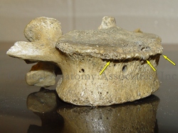
Spondylosis is an osteoarthritic degeneration of the vertebrae and the spine characterized by abnormal bony growths  on the vertebrae that can impinge on nerves and other structures causing pain and mobility problems. The definition of spondylosis also includes degenerative changes in the intervertebral discs.

The abnormal growth of portions of the vertebral body, usually forms "bone spurs", also referred to as "spondylophytes". The accompanying image shows a lumbar vertebra with spondylophytes. Click on the image for a larger depiction.

Spondylophytes on a lumbar vertebraImage property of: CAA.Inc. Photographer: David M. Klein
Back to MTD Main Page Subscribe to MTD

Rudolf Nissen, MD


This article is part of the series "A Moment in History" where we honor those who have contributed to the growth of medical knowledge in the areas of anatomy, medicine, surgery, and medical research.To search all the articles in this series, click here.
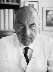
Dr. Rudolf Nissen (1896 - 1981).  Dr Nissen’s life is extraordinary. Born in the city of Neisse, Germany in 1896, he was the son of a local surgeon. He studied medicine in the Universities of Munich, Marburg, and Breslau. He was the pupil of the famous pathologist Albert Aschoff (discoverer of the heart’s AV node, along with Sunao Tawara).

Nissen became a professor of surgery in Berlin, and in 1933 moved to Turkey where he was placed in charge of the Department of Surgery of the University of Istanbul. In 1939 he moved to the US, first to the Massachusetts General Hospital and later to the Jewish Hospital in Brooklyn, New York. After becoming a US citizen, he moved again in 1952 to Basel, Switzerland as Chief of the Department of Surgery, where he retired in 1967. He died in 1981.

His contributions to surgery are innumerable. He wrote over 30 books and 450 journal articles. Known for the development in 1956 of what is today known as the “Nissen fundoplication” for esophageal hiatus hernia surgery, Nissen also worked with his assistant, Dr. Mario Rossetti to develop the “floppy Nissen fundoplication”, also known as the “Nissen-Rossetti procedure”. This would be enough to honor this man, still, he (with Sauerbruch) performed the first lung lobectomy and the first pneumonectomy (called then a total pneumonectomy). In 1949 he performed the first esophagectomy with a gastroesophagostomy for lower esophageal cancer.

Dr. Rudolph NissenOriginal imagecourtesy of Universit?t Basel.
His personal life is even more interesting. Drafted at 20, he fought in WWI and was wounded several times. In 1933, under the Nazi regime, he was ordered to fire all the Jewish-German assistants under his care. Being Jewish himself, he was told that he would keep his job, Nissen could not take this. He resigned his position and moved out of Germany.

Another little known fact is that he operated on Albert Einstein in 1948. He operated on Einstein because of intestinal cysts. Having found a developing abdominal aortic aneurysm, he reinforced it with cellophane, undoubtedly giving his patient a few extra years to live. Einstein died in 1955.

As a personal side note, our good friend Dr. Aaron Ruhalter scrubbed in with Dr. Nissen while serving as a surgical resident at the Brooklyn Jewish Hospital!

Sources:
1. “Rudolf Nissen: The man behind the fundoplication” Schein et al. Surgery 1999;125:347-53
2. “Rudolf Nissen (1896–1981)-Perspective” Liebermann-Meffert, D. J Gastrointest Surg (2010) 14 (Suppl 1):S58–S61
3. “The Life of Rudolf Nissen: Advancing Surgery Through Science and Principle” Fults, DW; Taussky, P. World J Surg (2011) 35:1402–1408
4. “Total Pneumonectomy” Nissen, R. Ann Thorac Surg 1980; 29:390-394
5. “Historical Development of Pulmonary Surgery” Nissen, R. Am J Surg 80: Jan 1955 9- 15

Back to MTD Main Page Subscribe to MTD

-oma

The suffix [-oma] means "tumor", "mass", or "growth". It should be noted that the word [tumor] is originally Latin, and means "swelling" or "bulging". Sometimes the plural form [-omata] can be used.

It is a general misconception that the suffix [-oma] or the term [tumor] are synonymous with "cancer". This is not so. [Tumor] only means a mass and the type of mass, benign or malignant is not implied in the term. A cancerous mass will be denoted by the addition of the root term [-carcin-] meaning "cancer" therefore the combined root and suffix will be [-carcinoma]. There are other root-suffix combinations that also mean "cancerous".

The suffix [-oma] can be found in many medical words, such as:

Hematoma: from the Greek root [-hem-] meaning "blood". A mass of blood
Myoma:  from the root [-my-] meaning "muscle". A mass, growth, or tumor of muscle.
Fibroma: from the root [-fibr-] meaning "fiber". A mass of fibers. 
Fibromyoma: the combining form of "muscle" is [myo-], therefore "a mass of fibers and muscle" (Pl. fibromyomata)
Adenoma: from the Greek [aden-] meaning "gland". A mass that has a glandular look to it or a mass in a gland 

Sources:
1. "The Language of Medicine" John H. Dirckx Pub: Harper & Row 1976
2. "Medical Meanings" Haubrich, William S. Am Coll Phys Philadelphia 1997
3. "The origin of Medical Terms" Skinner, AH, 1970

Back to MTD Main Page Subscribe to MTD

Giovanni Battista Morgagni


This article is part of the series "A Moment in History" where we honor those who have contributed to the growth of medical knowledge in the areas of anatomy, medicine, surgery, and medical research.To search all the articles in this series, click here.

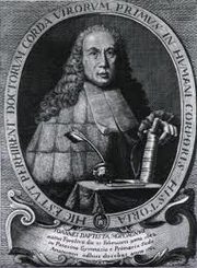
Giovanni Battista Morgagni (1682 - 1771). Italian anatomist, physician, and pathologist, Morgagni was born in the city of Forli. He started his medical studies at the University of Bologna, graduating in 1701 with a degree in Medicine and Philosophy. In 1712 he became a professor of anatomy at the University of Padua, Italy, 175 years after Andreas Vesalius. Morgagni was offered and accepted the Chair of Anatomy in 1715 at the University of Padua. Although Morgagni held a position at the anatomy department of the University of Padua, his name is associated mostly with his pathological studies.

Morgagni was interested in the works of Theophile Boneti (1620 - 1689), who started analyzing the correlation between post-mortem anatomical findings and diseases. He tried to establish a relation between the disease and the cause of death. In 1761 Morgagni published his most influential work "De Sedibus et Causis Morburum Per Anatomen Indagatis"  (On the Sites and Causes of Diseases, Investigated by Dissection). His work was essential for pathological anatomy to be recognized as a science in itself.

Giovanni Batista Morgagni
Morgagni was elected to become a member of several Academies of Science and Surgery: The Royal Society of London, The Academy of Science in Paris, The Berlin Academy of Science, and the Imperial Academy of Saint Petersburg in Russia. He is remembered today by several eponyms in anatomy and pathology:

• Morgagni's caruncle or lobe, referring to the miidle lobe of the prostate
• Morgagni's columns: the anal (or anorectal) colums
• Morgagni's concha, referring to the superior nasal concha
• Morgagni's foramina: two hiatuses in the respiratory diaphragm allowing for passage of the superior epigastric vessels
 Morgagni's hernia: an hiatal hernia through Morgagni's foramen, in the respiratory diaphragm
• Morgagni's ventricle: an internal pouch or dilation between the true and false vocal cords in the larynx
• Morgagni's nodules: the nodules at the point of coaptation of the leaflets (cusps) of the pulmonary valve. Erroneously called the "nodules of Arantius", which are only found in the aortic valve

Sources:
1. "A Note From History:The First Printed Case Reports of Cancer" Hadju, S.I. Cancer 2010;116:2493–8
2. "Giovanni Battista Morgagni" Klotz, O. Can Med Assoc J 1932 27:3 298-303
3. "Morgagni (1682 -1771)" JAMA 1964 187:12 948-950
Original imagecourtesy of National Institutes of Health.

Back to MTD Main Page Subscribe to MTD

Lumen

The term [lumen] is Latin and means "light". It is an international measure of light intensity. The plural form is either [lumens] or the Latin version [lumina].

The question is how did this term end up meaning the "internal opening or cavity of a hollow organ"? Dr. J. Dirckx (1976) explains that the term [lumen] in gastrointestinal anatomy was not used until the late 19th century, but it was initially used by microscopists looking at cross sections of small vessels. Since the empty interior of the vessel was represented on the histological slide as a clear, lighted region, they started calling that the "light" of the vessel, or [lumen].

The root term for [lumen] is [-lumin-] and can be found in over 30 words in English and related to light, such as luminosity, luminaria, luminescent, luminous, illumination,  illuminator, intraluminal, transluminal, endoluminal, etc.

The correct adjective form for [lumen] is [luminal], as stated by Haubrich (1997). The point is that the use of the term "lumenal" as an adjective form for lumen is not correct. This term can be seen in many textbooks today. The proper form for the NOTES acronym should be "Natural Orifice Transluminal Endoscopic Surgery" and not "translumenal". 

See how you can "illuminate" this article by hovering your cursor over the light bulb!

Sources:
1. "The Language of Medicine" John H. Dirckx Pub: Harper & Row 1976
2. "Medical Meanings" Haubrich, William S. Am Coll Phys Philadelphia 1997
3. "The origin of Medical Terms" Skinner, AH, 1970

Back to MTD Main Page Subscribe to MTD