Medical Terminology Daily (MTD) is a blog sponsored by Clinical Anatomy Associates, Inc. as a service to the medical community. We post anatomical, medical or surgical terms, their meaning and usage, as well as biographical notes on anatomists, surgeons, and researchers through the ages. Be warned that some of the images used depict human anatomical specimens.

You are welcome to submit questions and suggestions using our "Contact Us" form. The information on this blog follows the terms on our "Privacy and Security Statement" and cannot be construed as medical guidance or instructions for treatment.


We have 55 guests online


A Moment in History

Jean-Louis Petit

Jean Louis Petit
(1674 – 1750)

French surgeon and anatomist, Jean Louis Petit was born in Paris in on March 13, 1674.  His family rented an apartment at his house to Alexis Littre (1658 – 1726), a French anatomist. Petit became an apprentice of Littre at seven years of age, helping him in the dissections for his lectures and at an early age became the assistant in charge of the anatomic amphitheater.

Because of Petit’s dedication to anatomy and medicine, in 1690 at the age of sixteen, became a disciple of a famous Paris surgeon, Castel.

In 1692, Petit entered the French army and performed surgery in two military campaigns. By 1693 he started delivering lectures and was accepted as a great surgeon, being invited to the most difficult operations.  In 1700 he was appointed Chief Surgeon of the Military School in Paris and in the same year he received the degree of Master of Surgery from the Faculty of Paris.

In 1715 he was made a member of the Royal Academy of Sciences and an honorary member of the Royal Society of London. He was appointed by the King as the first Director General of the Royal Academy of Surgery when it was founded in 1731.

Petit’s written works are of historical importance.  “Traite des Maladies des Os” ( A Treatise on Bone Diseases);  “Traite des Maladies Chirurgicales et des Operation” (A Treatise on Surgical Diseases and their Operations” This last book was published posthumously in 1774. He also published a monograph on hemorrhage, another on lachrymal fistula, and others.

He was one of the first to perform choIecystotomy and mastoidotomy. His original tourniquet design for amputations saved many in the battlefield and the design of the same surgical instrument today has not changed much since its invention by him.

His name is remembered in the lumbar triangle, also called the "triangle of Petit", and the abdominal hernia that can ensue through that area of weakness, the lumbar hernia or "Petit's hernia".

Sources:
1. “Jean Louis Petit – A Sketch of his Life, Character, and Writings” Hayne, AP San Fran Western Lancet 1875 4: 446-454
2. “Oeuvres compl?tes de Jean-Louis Petit” 1837 Imprimerie de F. Chapoulaud
3. Extraits de l'eloge de Jean-Louis Petit Ius dans Ia seance publique de I' Academie royale de chirurgie du 26 mai 1750” Louis A. Chirurgie 2001: 126 : 475- 81


 "Clinical Anatomy Associates, Inc., and the contributors of "Medical Terminology Daily" wish to thank all individuals who donate their bodies and tissues for the advancement of education and research”.

Click here for more information


abebooks banner

bookplateink.com


Thebesian valve

UPDATED: The eponymic [Thebesian valve], or valve of the coronary sinus is a fold of endocardial tissue situated at the exit ostium of the coronary sinus. Named after Adam Christian Thebesius, the morphology of the valve presents with anatomical variations that go from total absence (see image here) to endocardial folds that cover up to 65% of the coronary sinus opening (see image in this article). The variations of the Thebesian valve include fenestrations, cribiriform valves, and the presence of pectineal cardiac muscle.

The Thebesian valve is important because it can be an obstruction to the passage of a catheter performing retrograde cardioplegia in electrophysiological studies, catheter ablation, and percutaneous mitral valve repair.

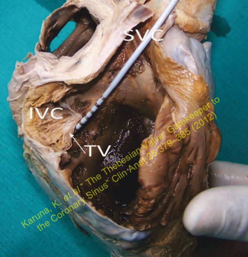
Thanks to Dr. Karuna Katti for allowing us the use of the image in this article. The image shows the interior of the right atrium with the inferior vena cava removed to demonstrate the Thebesian valve. In this case, the Thebesian valve covers >65% of the coronary sinus ostium. A catheter is being passed from the superior vena cava into the coronary sinus ostium.

Source:"The Thebesian Valve: Gatekeeper to the Coronary Sinus" Karuna Katti and Nikhil Prakash Patil Clin Anat 25:379–385 (2012)

IVC, inferior vena cava; SVC, superior vena cava; TV, Thebesian valve

IVC, inferior vena cava; SVC, superior vena cava; TV, Thebesian valve.
Click on the picture for a larger image 

MTD Main Page Subscribe to MTD