Medical Terminology Daily (MTD) is a blog sponsored by Clinical Anatomy Associates, Inc. as a service to the medical community. We post anatomical, medical or surgical terms, their meaning and usage, as well as biographical notes on anatomists, surgeons, and researchers through the ages. Be warned that some of the images used depict human anatomical specimens.

You are welcome to submit questions and suggestions using our "Contact Us" form. The information on this blog follows the terms on our "Privacy and Security Statement" and cannot be construed as medical guidance or instructions for treatment.


We have 184 guests online


A Moment in History

Jean-Louis Petit

Jean Louis Petit
(1674 – 1750)

French surgeon and anatomist, Jean Louis Petit was born in Paris in on March 13, 1674.  His family rented an apartment at his house to Alexis Littre (1658 – 1726), a French anatomist. Petit became an apprentice of Littre at seven years of age, helping him in the dissections for his lectures and at an early age became the assistant in charge of the anatomic amphitheater.

Because of Petit’s dedication to anatomy and medicine, in 1690 at the age of sixteen, became a disciple of a famous Paris surgeon, Castel.

In 1692, Petit entered the French army and performed surgery in two military campaigns. By 1693 he started delivering lectures and was accepted as a great surgeon, being invited to the most difficult operations.  In 1700 he was appointed Chief Surgeon of the Military School in Paris and in the same year he received the degree of Master of Surgery from the Faculty of Paris.

In 1715 he was made a member of the Royal Academy of Sciences and an honorary member of the Royal Society of London. He was appointed by the King as the first Director General of the Royal Academy of Surgery when it was founded in 1731.

Petit’s written works are of historical importance.  “Traite des Maladies des Os” ( A Treatise on Bone Diseases);  “Traite des Maladies Chirurgicales et des Operation” (A Treatise on Surgical Diseases and their Operations” This last book was published posthumously in 1774. He also published a monograph on hemorrhage, another on lachrymal fistula, and others.

He was one of the first to perform choIecystotomy and mastoidotomy. His original tourniquet design for amputations saved many in the battlefield and the design of the same surgical instrument today has not changed much since its invention by him.

His name is remembered in the lumbar triangle, also called the "triangle of Petit", and the abdominal hernia that can ensue through that area of weakness, the lumbar hernia or "Petit's hernia".

Sources:
1. “Jean Louis Petit – A Sketch of his Life, Character, and Writings” Hayne, AP San Fran Western Lancet 1875 4: 446-454
2. “Oeuvres compl?tes de Jean-Louis Petit” 1837 Imprimerie de F. Chapoulaud
3. Extraits de l'eloge de Jean-Louis Petit Ius dans Ia seance publique de I' Academie royale de chirurgie du 26 mai 1750” Louis A. Chirurgie 2001: 126 : 475- 81


 "Clinical Anatomy Associates, Inc., and the contributors of "Medical Terminology Daily" wish to thank all individuals who donate their bodies and tissues for the advancement of education and research”.

Click here for more information


abebooks banner

bookplateink.com


Atheroma

The medical word [atheroma], has the root term [-ather-] arising from the Greek [ath?ra] meaning "gruel", "porridge", or "groats". This refers to the consistency of the content of a soft atheromatous plaque. The suffix [-oma] means "mass", "growth" or "tumor". A mass of soft gruel-like substance. The plural form for atheroma is [atheromata].

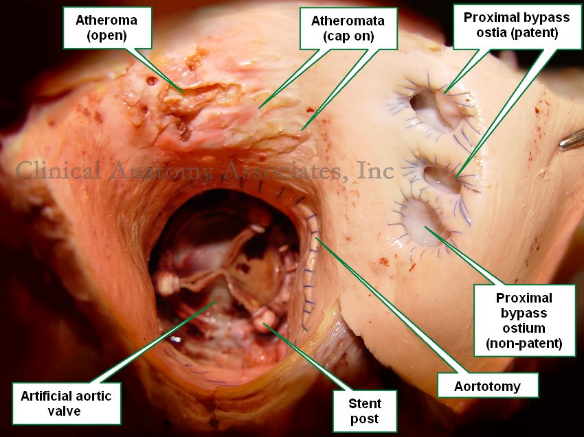
An atheroma is abnormal edema and accumulation of cholesterol and fatty acids with varying amount s of macrophages, fibroblasts and connective tissue in the tunica intima of an artery. It is usually covered by a “cap” of thicker, drier, yellowish fibrous material. An atheroma is a cavity filled with a fatty gruel-like material covered by a cap. Atheromatous disease is characterized by a large number of these masses in the walls of the arteries of a patient.

Superior view of the ascending aorta
Image property of: CAA.Inc. Photographer: D. M. Klein
Atheromata are found in smaller caliber arteries can reduce the lumen of the artery leading to ischemia and in the case of the coronary arteries, myocardial infarction.

The cap in an atheroma can be dislodged by the arterial blood flow in which case the content of the atheroma is emptied into the bloodstream becoming a fatty embolus. Since arteries become arterioles and then capillaries, this fatty embolus will flow distally to the point where it will lodge, blocking blood flow.

WARNING: The image of the pathology in this article is quite descriptive

The accompanying image is a clear depiction of this situation. This is a superior view of the ascending aorta. The patient in this case had an artificial aortic valve implanted and the aortotomy performed for the procedure is also indicated. The patient also had three coronary bypass grafts, one of which was clogged or non-patent. There are at least two atheromata with the cap still on. The image also shows at least one atheroma empty. This indicates that the content of the atheroma became a fatty embolus. Click on the image for a larger depiction.

Back to MTD Main Page Subscribe to MTD