Tracheal bronchus (pig bronchus)
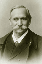
The tracheal bronchus is a known anatomical variation first described in 1785 by Eduard Sandifort (1742 – 1814), a Dutch physician and anatomist. Sandifort was the first to describe in detail what we know today as the Tetralogy of Fallot, although Nicolaus Steno (1638-1686) mentions the components of this pathology, he did not investigate it in detail as Sandifort did.
It is mostly known as the “pig bronchus” because of its similarity with pig anatomy; it is also called “bronchus suis”. When present, the tracheal bronchus usually arises on the right side of the trachea, about 2 cm. superior to the tracheal bifurcation (carina). The location of the bronchial aperture can vary between the cricoid cartilage of the larynx superiorly, and the tracheal bifurcation inferiorly.
The tracheal bronchus is usually the only airway supplying the upper right lobe of the lung, although it can share the airway with a smaller right upper lobe bronchus. In this case the tracheal bronchus is called “accessory tracheal bronchus”.
In cases where the origin of the tracheal bronchus is higher on the trachea, the trachea may present with distal stenosis. In rare cases, the tracheal bronchus may be found on the left side supplying air to the superior portion of the left lung.
The incidence of a tracheal bronchus varies between 1 to 5%, where the prevalence on the right side is 0.1 to 2% and 0.3 to 1% on the left side. It is sometimes found associated with other congenital abnormalities.
It is usually asymptomatic, but it can be found related to recurrent pulmonary infections, bronchiectasis, chronic bronchitis, and partial airway obstruction. It is most often discovered incidentally, usually during the intubation process for thoracic surgery.
Personal note: My thanks to Dr. Randall K. Wolf for suggesting this article.
Sources:
1. “Observationes anatomico-pathologicae” Sandifort, E. (1785). Lugduni Batavorum: Apud S. & J. Luchtmans.
2. “Congenital bronchial abnormalities revisited” Ghaye, B., Szapiro, D., Fanchamps, J. M., & Dondelinger, R. F. (2001). Radiographics, 21(1), 105–119.
3. “Tracheal bronchus: A rare cause of right upper lobe collapse” Ngernchuklin, P., Sumanac, K., & Behrsin, J. (2006).. Canadian Journal of Anesthesia, 53(12), 1227–1230
4. “Tracheal bronchus” radiopedia.org. Elfeky, M. 2023.
5: ”Bronchus suis – Case presentation” radiopedia.org. Ian Bickle.
Image modified from the original, public domain.


