Medical Terminology Daily (MTD) is a blog sponsored by Clinical Anatomy Associates, Inc. as a service to the medical community. We post anatomical, medical or surgical terms, their meaning and usage, as well as biographical notes on anatomists, surgeons, and researchers through the ages. Be warned that some of the images used depict human anatomical specimens.

You are welcome to submit questions and suggestions using our "Contact Us" form. The information on this blog follows the terms on our "Privacy and Security Statement" and cannot be construed as medical guidance or instructions for treatment.

We have 216 guests online


A Moment in History

Jean-Louis Petit

Jean Louis Petit
(1674 – 1750)

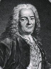
French surgeon and anatomist, Jean Louis Petit was born in Paris in on March 13, 1674.  His family rented an apartment at his house to Alexis Littre (1658 – 1726), a French anatomist. Petit became an apprentice of Littre at seven years of age, helping him in the dissections for his lectures and at an early age became the assistant in charge of the anatomic amphitheater.

Because of Petit’s dedication to anatomy and medicine, in 1690 at the age of sixteen, became a disciple of a famous Paris surgeon, Castel.

In 1692, Petit entered the French army and performed surgery in two military campaigns. By 1693 he started delivering lectures and was accepted as a great surgeon, being invited to the most difficult operations.  In 1700 he was appointed Chief Surgeon of the Military School in Paris and in the same year he received the degree of Master of Surgery from the Faculty of Paris.

In 1715 he was made a member of the Royal Academy of Sciences and an honorary member of the Royal Society of London. He was appointed by the King as the first Director General of the Royal Academy of Surgery when it was founded in 1731.

Petit’s written works are of historical importance.  “Traite des Maladies des Os” ( A Treatise on Bone Diseases);  “Traite des Maladies Chirurgicales et des Operation” (A Treatise on Surgical Diseases and their Operations” This last book was published posthumously in 1774. He also published a monograph on hemorrhage, another on lachrymal fistula, and others.

He was one of the first to perform choIecystotomy and mastoidotomy. His original tourniquet design for amputations saved many in the battlefield and the design of the same surgical instrument today has not changed much since its invention by him.

His name is remembered in the lumbar triangle, also called the "triangle of Petit", and the abdominal hernia that can ensue through that area of weakness, the lumbar hernia or "Petit's hernia".

Sources:
1. “Jean Louis Petit – A Sketch of his Life, Character, and Writings” Hayne, AP San Fran Western Lancet 1875 4: 446-454
2. “Oeuvres compl?tes de Jean-Louis Petit” 1837 Imprimerie de F. Chapoulaud
3. Extraits de l'eloge de Jean-Louis Petit Ius dans Ia seance publique de I' Academie royale de chirurgie du 26 mai 1750” Louis A. Chirurgie 2001: 126 : 475- 81


 "Clinical Anatomy Associates, Inc., and the contributors of "Medical Terminology Daily" wish to thank all individuals who donate their bodies and tissues for the advancement of education and research”.

Click here for more information


abebooks banner

bookplateink.com


-corp- / -corpor-

The root terms [-corp-] and [-corpor-] both arise from the Latin word [corpus] which means "body". The term corpus is used in human anatomy with the same meaning. Examples are:

• corpus vertebrale: The corpus or body of a vertebra

• corpus gastricum: The corpus or body of the stomach

• corpus callosum: A thick structure in the brain that communicates the cerebral hemispheres across the midline

The plural form is [corpora] as in the corpora cavernosa a pair of erectile bodies in the human genitalia.

In English both these roots can be found in words such as corpse, corporation, corporal, corpulent, corpuscle, etc.

Back to MTD Main Page Subscribe to MTD

Fundus

The word [fundus] is Latin and originally means "bottom", "base", or "foundation". The English root term [-found-] is derived from fundus in words such as "founder", "foundation", and "profound", as in "bottomless". Another derived root is [-fund-] with the meaning of "base capital".

Further evolution of the term lead to ancillary meaning of the term [fundus] as "pocket" as in the "bottom of a pocket". In fact, even today in Spanish, one of the word for "pocket" is "fundillo". This is the meaning that is used in human anatomy.

A fundus is a pocket-like segment or extension of an organ, such as the fundus of the stomach, the fundus of the gallbladder or the fundus of the uterus.

Back to MTD Main Page Subscribe to MTD

Eric Muhe


This article is part of the series "A Moment in History" where we honor those who have contributed to the growth of medical knowledge in the areas of anatomy, medicine, surgery, and medical research.To search all the articles in this series, click here.

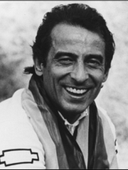
Prof. Dr. Eric Mühe  (1938 - 2005) Much has been written about who gets the glory of having performed the first laparoscopic cholecystectomy. Many famous names are in the annals of Surgery listed as pioneers in this field: Perissat, Berci, Mouret, Dubois, Sepulveda, Reddick, McKernan, Saye, Lizana, and others. Prof. Dr. Eric Mühe was usually not mentioned, but there is no doubt that on September 12, 1985, Dr. Mühe performed a laparoscopic cholecystectomy using a device of his own design (Galloscope) which lifted the anterior abdominal wall, maintained pneumoperitoneum, and doubled as a laparoscope. 

Dr. Mühe was an avid cyclist and built and repaired his own bycicles. He once  told me that he was looking at bicycle metal tube, and when looking through it, he thought that he could obtain access to the abdomen and the gallbladder with minimal changes. I am privileged to have known him and  I was sad to learn that he passed in 2005. Dr. Miranda

For more information in downloadable PDF format: CLICK HERE, or HERE

Prof Dr. Eric Muhe

Back to MTD Main Page Subscribe to MTD

Transverse processes

The [transverse processes] are two lateral bony processes found in most vertebrae, with the exception of the coccygeal vertebrae.

Transverse processes have regional variations. The transverse processes in the cervical vertebrae are heavily modified with anterior and posterior roots forming the boundaries of the foramina transversaria. They are "gutter-shaped" to accommodate the spinal nerves, and angled at 60 degrees anteriorly with a 15 degree inferior tilt.

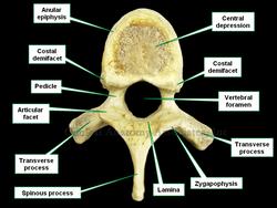
In the thoracic region the transverse processes point posterolaterally and present with an articular facet (costal facet) for the costal tubercle of a rib, forming the costotransverse joint.  The transverse processes of T11 and T12 do not have a costal facet.

Superior view of a thoracic vertebra
Image property of:CAA.Inc.Photographer:David M. Klein

The transverse processes in the lumbar region point slightly posterolateral, and they are larger. The longest transverse processes in the lumbar region are those of L3. The transverse processes of L5 are atyical in that they also point superolaterally at almost 45 degrees. This is because these transverse processes are attached to the posterior superior iliac spine and iliac crest by the iliolumbar ligament, contributing to the prevention of forward displacement of L5 over S1.

The sacral transverse processes contribute to the larger mass of the sacrum fusing to form the intermediate sacral crest on its posterior surface.

Thanks to Dr. Mary M Tuchscherer for her comments and improvement of this article.

Back to MTD Main Page Subscribe to MTD

Michael Servetus


This article is part of the series "A Moment in History" where we honor those who have contributed to the growth of medical knowledge in the areas of anatomy, medicine, surgery, and medical research.To search all the articles in this series, click here.

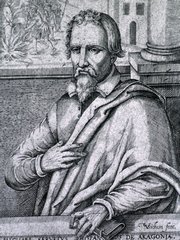
Michael Servetus (1511 -1553) was a Spanish theologian, physician, and anatomist. He is also known as  Miguel Servet, Miguel Serveto, and Michel de Villeneuve. Servetus had studies in a multitude of fields, including catography, mathematics, pharmacology, astronomy, etc. He was born in 1511 in Aragon, Spain. Servetus started his studies in law in 1531and Medicine in 1536, where he excelled as an anatomist. Just as Andreas Vesalius and William Harvey, he clashed with the Galenic vision of anatomy and physiology.  He correctly stated the theory of pulmonary circulation, but with no logical proof as Harvey.

Servetus was openly critical of the catholic church, publishing three books that openly questioned the Holy Trinity dogma. Servetus published his findings on pulmonary circulation in a controversial book "Cristianismi Restitutio", where pulmonary circulation was only one of the points he made which were mostly his position on the Holy Trinity and questioning the idea that everyone was predestined, as the catholic church professed at that time. His anatomical views were the least of his problems; because of this open criticism of Galen and the church. Servetus was burnt at the stake in Geneva on October 27, 1553.

Original image courtesy of National Library of Medicine.

 Michael Severtus

Back to MTD Main Page Subscribe to MTD

vertebral endplate

The [vertebral endplate] is the term used to denote a structure formed by the superior (and inferior)  aspect of the vertebral body and a layer of hyaline cartilage related to it. The vertebral endplate is formed by the cortical bone of the anular epiphysis, the central depression and the hyaline cartilage that fills the depresssion.

The vertebral endplate is important for the health of the intervertebral disk (IVD). Since the IVD loses its blood supply early in life all the nutrition to the IVD has to pass through the cortical bone and the hyaline cartilage. Note that hyaline cartilage is avascular. Separation of the components of the vertebral endplate due to trauma or other pathology can cause the IVD to become pathological. 

The vertebral endplate also plays an important role in the biomechanics of spine movement. Being slightly incurved, the endplate acts as a shock absorber, bending up to 1/2 mm. Damage to the endplate can reduce this function and set the stage for failure of the endplate and a potential vertebral fracture.

Superior view of a thoracic vertebra

Image property of:CAA.Inc.Photographer:David M. Klein

Sometimes the IVD can herniate through the endplate causing what is known as "Schmorl's bodies".


Back to MTD Main Page Subscribe to MTD