| Mondino de Luzzi (ca.1270 – 1326). Italian anatomist, born Raimondo de Luzzi in the city of Bologna circa 1270. He was also known as Mondino, Remondino, or Mundinus de Leutiis, de Lentiis, de Lucci, and other variations of his name. His father Nerino Franzoli was an apothecary, and Mondino also started working as such.
In 1290 he enrolled in the Medical School at the University of Bologna obtaining his medical degree circa 1290. Mondino stayed at the university, where he continued to teach until his death in 1326.
His major publication is “Anothomia Corporis Humani”, written circa 1316 and found only in manuscript form. It was finally printed in movable type in 1478, making it easily available to the public. While some authors like Singer, 1925 contend that this is his only publication, others discuss the possibility that Mondino de’ Luzzi wrote other books that have been adjudicated to other authors as at the time the name “Mondino” was very common.
“Anothomia Corporis Humani” is the first anatomical book based on actual dissections, and the book was organized almost as a dissection manual, explaining dissection techniques to visualize specific structures. Initially this book had no illustrations, but some were added in later publications.
|
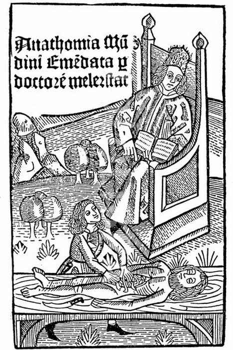
Title page of Anathomia Corporis Humanis by Mondino de Luzzi
|
| With over 40 editions, the last one in 1668, this book was used for almost 250 years. Mondino restarted human dissections in medical schools almost 1,500 years the medical school of Alexandria, leading many to call Mondino the “restorer of anatomy”.
It is said that Leonardo da Vinci (1452 – 1519) used one of Mondino’s books as a dissection manual to guide his own. Because Mondino followed Galen’s dictums and teachings, he was harshly criticized for his errors by Andreas Vesalius (1514 – 1564).
Although it is not clear if Mondino himself performed the actual dissections (he says he did), it is clear that he directed them. We know of two of his assistants: Otto Agenio Lustrolanus and Alessandra Giliani, the first woman prosector and anatomist. When Mondino died the same year as Alessandra Giliani, the expectation was that his assistant would continue the work of the master. Sadly Otto Agenio Lustrolanus died before he was 30 years old.
In the introduction to “Anothomia” Mondino says: "A work upon any science or art-as saith Galen-is issued for three reasons: First, that one may satisfy his friends. Second, that he may exercise his best mental powers. Third, that he may be saved from the oblivion incident to old age. Therefore, moved by these three causes, I have proposed to my pupils to compose a certain work on Medicine.”
"And because a knowledge of the parts to be subjected to medicine (which is the human body, and the names of its various divisions) is a part of medical science, as saith Averrhoes in his first chapter, in the section on the definition of medicine, for this reason among others I have set out to lay before you the knowledge of the parts of the human body which is derived from anatomy, not attempting to use a lofty style, but the rather that which is suitable to a manual procedure."
Sources:
1. “Mondino de' Luzzi's commentary on the Canones Generales of Mesue the Younger” Welborn, MC. Isis , 22: 1 (1934) , 8-11
2. “Medieval neuroanatomy: the text of Mondino dei Luzzi and the plates of Guido da Vigevano” Orly R. J Hist Neurosci. 1997 6 (2):113-123
3. “Mondino de Luzzi (1270-1326) Restaurador de la Disecci?n Anat?mica” Rever?n, RR. Informe Medico 2007; 9 (12):589-592
4. “The history and illustration of anatomy in the Middle Ages” Gurunluoglu, R, et al. J Med Biogr 2013 21: 219 – 229
5. “The Mondino Myth” Pilcher, LS. 1906
Original image courtesy of NLM
|


