Medical Terminology Daily (MTD) is a blog sponsored by Clinical Anatomy Associates, Inc. as a service to the medical community. We post anatomical, medical or surgical terms, their meaning and usage, as well as biographical notes on anatomists, surgeons, and researchers through the ages. Be warned that some of the images used depict human anatomical specimens.

You are welcome to submit questions and suggestions using our "Contact Us" form. The information on this blog follows the terms on our "Privacy and Security Statement" and cannot be construed as medical guidance or instructions for treatment.


We have 189 guests online


A Moment in History

Jean-Louis Petit

Jean Louis Petit
(1674 – 1750)

French surgeon and anatomist, Jean Louis Petit was born in Paris in on March 13, 1674.  His family rented an apartment at his house to Alexis Littre (1658 – 1726), a French anatomist. Petit became an apprentice of Littre at seven years of age, helping him in the dissections for his lectures and at an early age became the assistant in charge of the anatomic amphitheater.

Because of Petit’s dedication to anatomy and medicine, in 1690 at the age of sixteen, became a disciple of a famous Paris surgeon, Castel.

In 1692, Petit entered the French army and performed surgery in two military campaigns. By 1693 he started delivering lectures and was accepted as a great surgeon, being invited to the most difficult operations.  In 1700 he was appointed Chief Surgeon of the Military School in Paris and in the same year he received the degree of Master of Surgery from the Faculty of Paris.

In 1715 he was made a member of the Royal Academy of Sciences and an honorary member of the Royal Society of London. He was appointed by the King as the first Director General of the Royal Academy of Surgery when it was founded in 1731.

Petit’s written works are of historical importance.  “Traite des Maladies des Os” ( A Treatise on Bone Diseases);  “Traite des Maladies Chirurgicales et des Operation” (A Treatise on Surgical Diseases and their Operations” This last book was published posthumously in 1774. He also published a monograph on hemorrhage, another on lachrymal fistula, and others.

He was one of the first to perform choIecystotomy and mastoidotomy. His original tourniquet design for amputations saved many in the battlefield and the design of the same surgical instrument today has not changed much since its invention by him.

His name is remembered in the lumbar triangle, also called the "triangle of Petit", and the abdominal hernia that can ensue through that area of weakness, the lumbar hernia or "Petit's hernia".

Sources:
1. “Jean Louis Petit – A Sketch of his Life, Character, and Writings” Hayne, AP San Fran Western Lancet 1875 4: 446-454
2. “Oeuvres compl?tes de Jean-Louis Petit” 1837 Imprimerie de F. Chapoulaud
3. Extraits de l'eloge de Jean-Louis Petit Ius dans Ia seance publique de I' Academie royale de chirurgie du 26 mai 1750” Louis A. Chirurgie 2001: 126 : 475- 81


 "Clinical Anatomy Associates, Inc., and the contributors of "Medical Terminology Daily" wish to thank all individuals who donate their bodies and tissues for the advancement of education and research”.

Click here for more information


abebooks banner

bookplateink.com


Iatrogenic

This medical term is Greek and is composed of [?ατρός] (iatros) meaning "doctor", "physician", or "healer" and the suffix [-(o)genic], meaning "creation", "born of", or "beggining". An iatrogenic condition is that which is caused or created by the doctor or the hospital.

This is an expensive word, as iatrogenic conditions may lead to a lawsuit!

Back to MTD Main Page Subscribe to MTD

Leiomyoma

The word [leiomyoma] is of Greek origin with combined root terms. The term [-lei(o)-] arises from the Greek [λείος] meaning "smooth", the other root is [μυς] (mys) meaning "muscle". The suffix [-oma] means "tumor" or "mass". A [leiomyoma] is a "smooth muscle tumor". The medical plural form is [leiomyomata], or it can be [leiomyomas].

Since smooth muscle is involuntary muscle, leiomyomata are usually found in viscera. The most common leiomyomata are found in the uterus (see image), in the muscular or submucosal layer of the digestive system, mostly jejunum and ileum, and gallbladder, or in smooth muscle of the skin. The term itself does not imply that leiomyomata are cancerous, and most leiomyomata are not.

Uterine leiomyoma (www.wikipedia.org)Original image courtesy of www.Wikipedia.org

Back to MTD Main Page Subscribe to MTD

Mesoappendix

Terminal ileum, cecum, and vermiform appendix. Public domain
Terminal ileum, cecum,
and vermiform appendix

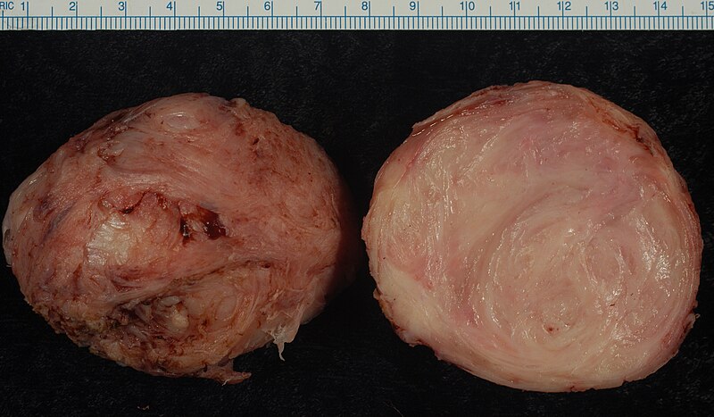
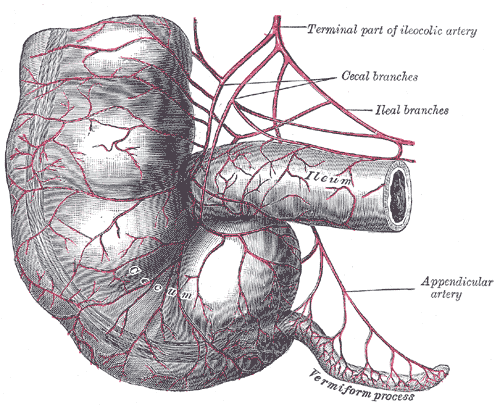
The [mesoappendix] is a triangular-shaped double-layered peritoneal membrane related to the vermiform appendix. One of the sides attaches to the vermiform appendix, the other is free, and the third one attaches to the ileum and the cecum. This last attachment varies in extension, giving the cecum varying degrees of mobility.

The mesoappendix contains the appendicular artery. This artery arises either from the ileocolic artery or the from the posterior ileocecal artery. The mesoappendix also contains the appendicular veins, lymphatics, lymphatic nodes, and fat.

In the female, there can be an extension of the mesoappendix that communicates with the broad ligament of the uterus. It is called the appendiculoovarian ligament, or Clado's ligament. This ligament may contain the appendiculoovarian artery, an anastomosis between the appendicular artery and the ovarian artery. The lymphatics contained in this appendiculoovarian ligament can also establish a lymphatic communication between the ovary and the vermiform appendix.

Sources:
1 "Tratado de Anatomia Humana" Testut et Latarjet 8 Ed. 1931 Salvat Editores, Spain
2. "Anatomy of the Human Body" Henry Gray 1918. Philadelphia: Lea & Febiger Image modified by CAA, Inc. Original image by Henry Vandyke Carter, MD., courtesy of bartleby.com


Alessandra Giliani


This article is part of the series "A Moment in History" where we honor those who have contributed to the growth of medical knowledge in the areas of anatomy, medicine, surgery, and medical research.To search all the articles in this series, click here.
Alessandra Giliani (1307 – 1326). Italian prosector and anatomist. Alessandra Giliani is the first woman to be on record as being an anatomist and prossector. She was born on 1307 in the town of Persiceto in northern Italy.

She was admitted to the University of Bologna circa 1323. Most probably she studied philosophy and the foundations of anatomy and medicine. She studied under Mondino de Luzzi (c.1270 – 1326), one of the most famous teachers at Bologna.

Giliani was the prosector for the dissections performed at the Bolognese “studium” in the Bologna School of Anatomy. She developed a technique (now lost to history) to highlight the vascular tree in a cadaver using fluid dyes which would harden without destroying them. Giliani would later paint these structures using a small brush. This technique allowed the students to see even small veins.

Giliani died at the age of 19 on March 26, 1326, the same year that her teacher Mondino de Luzzi died.  It is said that she was buried in front of the Madonna delle Lettere in the church of San Pietro e Marcellino at the Hospital of Santa Maria del Mareto in Florence by Otto Agenius Lustrulanus, another assistant to Modino de Luzzi.

Title page of Anathomia Corporis Humanis by Mondino de Luzzi. Image courtesy of the National Library of Medicine
Title page of Anathomia Corporis Humanis by Mondino de Luzzi
Some ascribe to Agenius a love interest in Giliani because of the wording of the plaque that is translated as follows:

"In this urn enclosed are the ashes of the body of
Alessandra Giliani, a maiden of Persiceto.
Skillful with her brush in anatomical demonstrations
And a disciple equaled by few,
Of the most noted physician, Mondino de Luzzi,
She awaits the resurrection.
She lived 19 years: She died consumed by her labors
March 26, in the year of grace 1326.
Otto Agenius Lustrulanus, by her taking away
Deprived of his better part, inconsolable for his companion,
Choice and deservinging of the best from himself,
Has erected this plaque"

Sir William Osler says of Alessandra Giliani “She died, consumed by her labors, at the early age of nineteen, and her monument is still to be seen”

The teaching of anatomy in the times of Mondino de Luzzi and Alessandra Giliani required the professor to be seated on a high chair or “cathedra” from whence he would read an anatomy book by Galen or another respected author while a prosector or “ostensor” would demonstrate the structures to the student. The professor would not consider coming down from the cathedra to discuss the anatomy shown. This was changed by Andreas Vesalius.

The image in this article is a close up of the title page of Mondino’sAnothomia Corporis Humani” written in 1316, but published in 1478. Click on the image for a complete depiction of this title page. I would like to think that the individual doing the dissection looking up to the cathedra and Mondino de Luzzi is Alessandra Giliani… we will never know.

The life and death of Alessandra Giliani has been novelized in the fiction book “A Golden Web” by Barbara Quick.

Sources
1. “Books of the Body: Anatomical Ritual and Renaissance Learning” Carlino, A. U Chicago Press, 1999
2. “Encyclopedia of World Scientists” Oakes, EH. Infobase Publishing, 2002
3. “The Biographical Dictionary of Women in Science”Harvey, J; Ogilvie, M. Vol1. Routledge 2000
4. “The Evolution of Modern Medicine” Osler, W. Yale U Press 1921
5. “The Mondino Myth” Pilcher, LS. 1906
Original image courtesy of NLM

Back to MTD Main Page Subscribe to MTD

Peritoneum

The word [peritoneum] has a Greek origin [περίτόνοςαιον]. Loosely translated it has the preffix [peri-] meaning "around", the root [-ton-] from the Greek [tonos], meaning "to stretch", and the suffix [-eum] meaning "a membrane". It is "a membrane that is stretched around".

The peritoneum is a thin serosal membranous sac found in the abdominopelvic cavity. Histologically it is composed of a layer of mesothelium supported by a layer of connective tissue. Being a serosal sac, it contains in its interior a small amount of peritoneal fluid. The pathological accumulation of peritoneal fluid is called ascitis.

Although the peritoneum is one continuous membrane, and because of its relation to the organs and the abdominal wall, the peritoneum is described as formed by two components:

• Parietal peritoneum: The parietal peritoneum is that portion of the peritoneal sac related to or in contact with the walls of the abdomen and the pelvis.

• Visceral peritoneum: The visceral peritoneum is that portion of the peritoneal sac related to or in contact with the abdominopelvic viscera. I this case the peritoneum encases the viscera almost completely and is referred to as their serosa layer i,e: serosa layer of the ileum.

Abdominal contents (Testut Latarjet 1931)

The double-layered portions of the peritoneal sac that stretch between organs or between organs and the abdominal wall are known by different names. They can be called an abdominopelvic [ligament], a [mesentery], a [meso..(something)], or an [omentum]. These structures are covered in separate articles. Some of these structures are:

Falciform ligament: A sickle-shaped double fold of peritoneum related to the liver
Ligament of Treitz: Also known as the "suspensory ligament of the duodenum"
• Infundibulopelvic ligament: A fold of peritoneum containing the ovarian arteries and veins
• Lesser omentum: The lesser omentum is one of the two double-folds of peritoneum related to the stomach
• Mesosigmoid: A double peritoneal membrane related to the sigmoid colon
• Transverse mesocolon: A double peritoneal membrane related to the transverse colon
Mesoappendix: A double peritoneal membrane related to the vermiform appendix, etc.

Sources:
1. "Clinically Oriented Anatomy" Moore, KL. 3r Ed. Williams & Wilkins 1992
2. "The origin of Medical Terms" Skinner, AH, 1970
3. "Tratado de Anatomia Humana" Testut et Latarjet 8 Ed. 1931 Salvat Editores, Spain
Image modified from the original from Testut and Latajet, 1931. Click in the image for a larger depiction
Thanks to Dr. Randall Wolf for suggesting this article

Back to MTD Main Page Subscribe to MTD

Cervical

A derivate of the Latin root [cervix] or [cervicis] meaning "neck". The word [cervical] means "pertaining to the neck".

The term is used in many areas and structures of the human body:

• Cervical spine: refers to the spinal column region formed by the seven cervical vertebrae. See image

• Uterine cervix: The inferior region of the uterus which projects partially into the vagina.

• Cervical rib: An anatomic variation where one or more supernumerary ribs are found related to the lower cervical vertebrae. This anomaly can cause clinical symptoms.

Images property of: CAA.Inc. Artist: Dr. E. Miranda

Cervical Vertebrae
Back to MTD Main Page Subscribe to MTD