Medical Terminology Daily (MTD) is a blog sponsored by Clinical Anatomy Associates, Inc. as a service to the medical community. We post anatomical, medical or surgical terms, their meaning and usage, as well as biographical notes on anatomists, surgeons, and researchers through the ages. Be warned that some of the images used depict human anatomical specimens.

You are welcome to submit questions and suggestions using our "Contact Us" form. The information on this blog follows the terms on our "Privacy and Security Statement" and cannot be construed as medical guidance or instructions for treatment.


We have 92 guests online


A Moment in History

Jean-Louis Petit

Jean Louis Petit
(1674 – 1750)

French surgeon and anatomist, Jean Louis Petit was born in Paris in on March 13, 1674.  His family rented an apartment at his house to Alexis Littre (1658 – 1726), a French anatomist. Petit became an apprentice of Littre at seven years of age, helping him in the dissections for his lectures and at an early age became the assistant in charge of the anatomic amphitheater.

Because of Petit’s dedication to anatomy and medicine, in 1690 at the age of sixteen, became a disciple of a famous Paris surgeon, Castel.

In 1692, Petit entered the French army and performed surgery in two military campaigns. By 1693 he started delivering lectures and was accepted as a great surgeon, being invited to the most difficult operations.  In 1700 he was appointed Chief Surgeon of the Military School in Paris and in the same year he received the degree of Master of Surgery from the Faculty of Paris.

In 1715 he was made a member of the Royal Academy of Sciences and an honorary member of the Royal Society of London. He was appointed by the King as the first Director General of the Royal Academy of Surgery when it was founded in 1731.

Petit’s written works are of historical importance.  “Traite des Maladies des Os” ( A Treatise on Bone Diseases);  “Traite des Maladies Chirurgicales et des Operation” (A Treatise on Surgical Diseases and their Operations” This last book was published posthumously in 1774. He also published a monograph on hemorrhage, another on lachrymal fistula, and others.

He was one of the first to perform choIecystotomy and mastoidotomy. His original tourniquet design for amputations saved many in the battlefield and the design of the same surgical instrument today has not changed much since its invention by him.

His name is remembered in the lumbar triangle, also called the "triangle of Petit", and the abdominal hernia that can ensue through that area of weakness, the lumbar hernia or "Petit's hernia".

Sources:
1. “Jean Louis Petit – A Sketch of his Life, Character, and Writings” Hayne, AP San Fran Western Lancet 1875 4: 446-454
2. “Oeuvres compl?tes de Jean-Louis Petit” 1837 Imprimerie de F. Chapoulaud
3. Extraits de l'eloge de Jean-Louis Petit Ius dans Ia seance publique de I' Academie royale de chirurgie du 26 mai 1750” Louis A. Chirurgie 2001: 126 : 475- 81


 "Clinical Anatomy Associates, Inc., and the contributors of "Medical Terminology Daily" wish to thank all individuals who donate their bodies and tissues for the advancement of education and research”.

Click here for more information


abebooks banner

bookplateink.com


Scalpel

The term [scalpel] arises from the Latin word [scalpellum], meaning “a small knife”. Celsus (c.25BC - c.50BC) in De Re Medicina, uses the term [scalpellus] to refer to a surgeon’s knike.

The use of knives in Medicine is very old. 10,000 year old Mesolithic skulls have been found with craniotomies made with flint stone knives. The first one to describe surgical knives in recorded history was Hippocrates of Cos (460 BC - 370 BC).

Surgical knives were developed in a multitude of shape and forms, some of them veritable works of art. These scalpels were made by cutlery makers on demand by surgeons and had a fixed blade that had to be sharpened constantly, as surgery requires a very sharp blade.

The invention of the disposable razor by King Gillette in 1904 changed this. In 1910, Dr. John Benjamin Murphy (1857 – 1916) invented a handle that could hold a single-sided or a double-sided safety razor blade.

The design was improved by the invention of a disposable, sterilized surgical blade that could be easily installed on a reusable, sterilizable metal handle. Today sterile, disposable handle-blade combination scalpels of varying shapes and sizes are available, ensuring the surgeon the sharpest scalpel every time.

Sources
1. “The surgical knife” Ochsner J. Tex Heart Inst J. 2009; 36(5): 441–443
2. "History of the Surgical Blade" Shuja, A. Indep Rev Oct-Dec 2012;14(10-12)
3. "Masters of the Scalpel: The History of Surgery"" Riedman SR, R MacNally 1962  

Images courtesy of 
Wikipedia

Different scalpel models (Wikipedia)
    Different scalpel models

Back to MTD Main Page Subscribe to MTD

Ureter

The [ureters] are two long, thin, bilateral muscular tubes that extend from the pelvis of the ipsilateral kidney to the posteroinferior aspect of the urinary bladder. The ureters are retroperitoneal structures and their function is the transport of urine between the kidney and the urinary bladder.

The word arises from the Greek [ουρητήρ], meaning "urinary duct". The term was originally used to denote both the urethra (in its singular form) and the ureters (in its plural form). Because of the problems using the term, the word [urethra] was created.

The ureter is composed of three layers: a mucosa, a thick muscular layer, and an adventitia. The muscular layer is itself composed of an deep longitudinal layer and a superficial circular layer formed by spiral smooth muscle fibers. In the distal portion of the ureter there is a third layer added to the ones mentioned, a thick longitudinal layer that extends into the walls of the urinary bladder. Since the ureter is a retroperitoneal organ, in the areas where the ureter is in contact with the parietal peritoneum, it can be said that the ureter presents with a fourth layer. The muscular construction of the ureters in layers allows for peristalsis that helps the flow of urine.

Longitudinal section of a kidney (wikipedia.org) 1-Renal pyramid, 3-Renal artery, 4-Renal vein, 5-Renal hilum, 6-Renal pelvis, 7- Ureter, 8-Minor calyx, 9-Renal capsule, 14-Minor calyx, 15- Major calyx, 16-Renal papilla, 17-Renal column
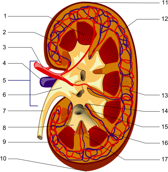
    Longitudinal section of a kidney. 1-Renal pyramid, 3-Renal artery, 4-Renal vein, 5-Renal hilum, 6-Renal pelvis, 7- Ureter, 8-Minor calyx, 9-Renal capsule, 14-Minor calyx, 15- Major calyx, 16-Renal papilla, 17-Renal column .

In the female the pelvic ureter passes just inferior to the uterine artery, as situation that surgeons refer to as "water under the bridge". Proper identification of the ureter is critical to avoid damage to this structure during a total hysterectomy. Surgeons will use the peristaltic movement of the ureter to identify it.

In its trajectory the ureters descend anterior to the psoas major muscle, pass over the pelvic brim, just anterior to the origin of the internal iliac artery, then hug the lateral pelvic wall and enter the urinary bladder from a posteroinferior aspect. As it descends the ureters receive blood supply from different arteries: renal arteries, gonadal (testicular or ovarian) arteries, aorta, common iliac arteries, and the inferior vesical arteries. The ureters also receive sympathetic and parasympathetic innervation from the autonomic nervous system renal plexus.

Sources
1. "Gray's Anatomy"38th British Ed. Churchill Livingstone 1995
2. "Tratado de Anatomia Humana" Testut et Latarjet 8 Ed. 1931 Salvat Editores, Spain
3. "The origin of Medical Terms" Skinner, AH, 1970
4. "Histology; a Text and Atlas" Ross MH 3rd Ed. Williams and Wilkins 1995
Images courtesy of 
Wikipedia

Back to MTD Main Page Subscribe to MTD

-mamm-

The root term [-mamm-arises from the Latin [mamma] (Pl. mammae), meaning "breast".  The synonymous term [-mast-] arises from the Greek. This prefix is used in medical terms such as:

• Mammal: An animal with breasts or that feeds trough breast milk

• Mammary: Pertaining to the breast

• Mammoplasty: The suffix [-(o)plasty] is used to mean "surgical reshaping". Surgical reshaping of the breast. Can also be referred to as a mastoplasty

• Mammogram: The suffix [-(o)gram] means "to record", or "to write". The results of a breast examination, usually a film obtained with imaging technology

• Mammography: The suffix [-(o)graphy] means "to record", or "to examine". A breast examination, usually with imaging technology

Brainstem. Anteroinferior view
Image property of: CAA.Inc. Photography: Efrain Klein

• Mammillary bodies: Two small, round structures found in the anterior aspect of the mesencephalon (midbrain) between the crura cerebri. See accompanying image. Click on the image for a larger depiction.

• Mammillary processes: Also known as the mammilary tubercles. These are small bony processes found in the transverse processes of the lumbar vertebrae

Back to MTD Main Page Subscribe to MTD

William T.G. Morton


This article is part of the series "A Moment in History" where we honor those who have contributed to the growth of medical knowledge in the areas of anatomy, medicine, surgery, and medical research.To search all the articles in this series, click here.
William Thomas Green Morton  (1819-1868). American dentist, was born in Charlton, MA. In 1840, He started his studies at the Baltimore College of Dental Surgery. Interestingly, he left college without graduating to study with Horace Wells, DDS, in Hartford, CT. He started Medical school at Harvard, and left without graduating. In 1852 Morton received his honorary MD degree.

In 1846, Morton was able to successfully and painlessly extract a tooth from a patient under ether administration. To this effect he used an ether inhaler of his own invention that he called “Letheon”. This led to his now-famous demonstration of the use of ether as an anesthetic on October 1846 at the Massachusetts General Hospital. This is the first-ever recorded use of anesthesia as a way to reduce or eliminate pain in surgery, one of the revolutions in surgery, the other being Lister’s antiseptic technique.

Morton tried, unsuccessfully, to establish a patent on his discovery against Horace Wells (1815 – 1848). The litigation costs led him to poverty and he died with no money on 1868.

William TG Morton
Although the American Dental Association in 1864 passed a formal resolution stating that Horace Wells was the discoverer of anesthesia, the name of William TG Morton has been tied to it since.

Sources
1. “William Thomas Green Morton (1819-1868” Keys. TE. Anesth and Analg (1973) 52: (2) 166
2. ” Morton, dentist, who first publicly demonstrated ether anesthesia; a short biography” Archer WH, William TG. J Am Dent Assoc. 1946 Dec 1;33(23):1528-32
3. “Horace Wells: Discoverer of Anesthesia” Jacobsohn PH. Anesth Prog (1995) 42:73-75
4. “The last days of William Thomas Green Morton” Vandam LD J Clin Anesth (1996) Sep;8(6):431-4334

Back to MTD Main Page Subscribe to MTD

Lung

A [lung] is one of the two main respiratory organs. It is an organ filled with minute air sacs or alveoli, giving its parenchyma the look and feel of a spongy tissue. Its function is the reoxygenation of blood.

Each lung is located on each side of the thoracic cavity, each one surrounded by a serosa membrane called "pleura". The pleura forms a sac that surrounds the lung and also lines the thoracic wall. The pleura that lines the thoracic wall is called "parietal pleura", while the pleura that lines the lung is called the "visceral pleura". An extension of the visceral pleura inferior to the pulmonary hilum forms the pulmonary ligament.

A lung has three surfaces and an apex. The inferior surface, base, or diaphragmatic surface; the medial surface or mediastinal surface, as if forms the lateral wall of the mediastinum; and the lateral or costal surface.

The lungs are also divided into lobes. The right lung has three lobes: superior, middle, and inferior while the left lung only has two lobes: superior and inferior. Because the heart has a left-sided tilt the left lung is slightly smaller than the right lung.

There are four structures that enter or leave the lung and they are found at the pulmonary hilum in the medial surface of the lung. These structures are: the main or primary bronchus, the pulmonary artery, and two pulmonary veins.

Medial surface of a left lung
Image property of: CAA.Inc.Photographer:David M. Klein
After entering the lung through the hilum each bronchus will divide into smaller and smaller bronchi eventually opening into microscopic air sacs called the alveoli. Each alveolus or alveolar sac is surrounded by capillaries. It is here where gas exchange takes place allowing for blood reoxygenation.

When examined, a lung will show the impressions of the organs that are in contact with it. Click on the accompanying image and you will be able to see the impressions left by the aorta and the heart

Back to MTD Main Page Subscribe to MTD

Atheroma

The medical word [atheroma], has the root term [-ather-] arising from the Greek [ath?ra] meaning "gruel", "porridge", or "groats". This refers to the consistency of the content of a soft atheromatous plaque. The suffix [-oma] means "mass", "growth" or "tumor". A mass of soft gruel-like substance. The plural form for atheroma is [atheromata].

An atheroma is abnormal edema and accumulation of cholesterol and fatty acids with varying amount s of macrophages, fibroblasts and connective tissue in the tunica intima of an artery. It is usually covered by a “cap” of thicker, drier, yellowish fibrous material. An atheroma is a cavity filled with a fatty gruel-like material covered by a cap. Atheromatous disease is characterized by a large number of these masses in the walls of the arteries of a patient.

Superior view of the ascending aorta
Image property of: CAA.Inc. Photographer: D. M. Klein
Atheromata are found in smaller caliber arteries can reduce the lumen of the artery leading to ischemia and in the case of the coronary arteries, myocardial infarction.

The cap in an atheroma can be dislodged by the arterial blood flow in which case the content of the atheroma is emptied into the bloodstream becoming a fatty embolus. Since arteries become arterioles and then capillaries, this fatty embolus will flow distally to the point where it will lodge, blocking blood flow.

WARNING: The image of the pathology in this article is quite descriptive

The accompanying image is a clear depiction of this situation. This is a superior view of the ascending aorta. The patient in this case had an artificial aortic valve implanted and the aortotomy performed for the procedure is also indicated. The patient also had three coronary bypass grafts, one of which was clogged or non-patent. There are at least two atheromata with the cap still on. The image also shows at least one atheroma empty. This indicates that the content of the atheroma became a fatty embolus. Click on the image for a larger depiction.

Back to MTD Main Page Subscribe to MTD