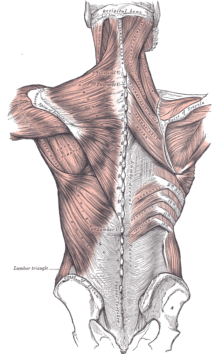
Posterior view of the superficial and
intermediate muscle layers of the back
The [trapezius] is a bilateral muscle belonging to the superficial muscles of the back. On each side it is a flat, thin triangular muscle that spans the neck, shoulders and the superior and middle aspect of the back. When seen together these two triangular muscles form a diamond-shaped quadrangle from which its name derives. The word originates in the Greek [τραπεζι] meaning "a four-legged table" (four sides). This word later evolved into the New Latin [trapezium].
In the midline the trapezius muscle attaches to the inion (external occipital protuberance), the ligamentum nuch?, the spinous processes of the seventh cervical vertebra (vertebra prominens), and the spinous processes of all the thoracic vertebr?.
The trapezius’ muscle fibers have three orientations. From the midline the superior fibers course inferolaterally to attach to the posterior border of the lateral third of the clavicle. The middle fibers course laterally to attach to the medial margin of the acromion, and posterior border of the spine of the scapula. The inferior fibers course superolaterally to attach to the spine of the scapula by way of an aponeurosis.
Because of their attachments, the superior and inferior fibers of the trapezius act coordinatedly to rotate the scapula, while the middle fibers act to retract the scapula. The superior fibers also act to slightly elevate the scapula. The trapezius muscle is sometimes described as an accessory respiratory muscle.
The trapezius muscle receives muscular innervation by way of the spinal accessory nerve (11th Cranial Nerve) which courses on the deep aspect of the muscle along with the superficial branch of the transverse cervical artery and vein. The muscle also receives sensory innervation by way of nerves arising from the ventral rami of the 3rd and 4th spinal nerves.
The trapezius is one of the 17 muscles that attach to the scapula.
Sources:
1 "Tratado de Anatomia Humana" Testut et Latarjet 8 Ed. 1931 Salvat Editores, Spain
2. "Anatomy of the Human Body" Henry Gray 1918. Philadelphia: Lea & Febiger
Original images courtesy of bartleby.com