Medical Terminology Daily (MTD) is a blog sponsored by Clinical Anatomy Associates, Inc. as a service to the medical community. We post anatomical, medical or surgical terms, their meaning and usage, as well as biographical notes on anatomists, surgeons, and researchers through the ages. Be warned that some of the images used depict human anatomical specimens.

You are welcome to submit questions and suggestions using our "Contact Us" form. The information on this blog follows the terms on our "Privacy and Security Statement" and cannot be construed as medical guidance or instructions for treatment.


We have 158 guests online


A Moment in History

Jean-Louis Petit

Jean Louis Petit
(1674 – 1750)

French surgeon and anatomist, Jean Louis Petit was born in Paris in on March 13, 1674.  His family rented an apartment at his house to Alexis Littre (1658 – 1726), a French anatomist. Petit became an apprentice of Littre at seven years of age, helping him in the dissections for his lectures and at an early age became the assistant in charge of the anatomic amphitheater.

Because of Petit’s dedication to anatomy and medicine, in 1690 at the age of sixteen, became a disciple of a famous Paris surgeon, Castel.

In 1692, Petit entered the French army and performed surgery in two military campaigns. By 1693 he started delivering lectures and was accepted as a great surgeon, being invited to the most difficult operations.  In 1700 he was appointed Chief Surgeon of the Military School in Paris and in the same year he received the degree of Master of Surgery from the Faculty of Paris.

In 1715 he was made a member of the Royal Academy of Sciences and an honorary member of the Royal Society of London. He was appointed by the King as the first Director General of the Royal Academy of Surgery when it was founded in 1731.

Petit’s written works are of historical importance.  “Traite des Maladies des Os” ( A Treatise on Bone Diseases);  “Traite des Maladies Chirurgicales et des Operation” (A Treatise on Surgical Diseases and their Operations” This last book was published posthumously in 1774. He also published a monograph on hemorrhage, another on lachrymal fistula, and others.

He was one of the first to perform choIecystotomy and mastoidotomy. His original tourniquet design for amputations saved many in the battlefield and the design of the same surgical instrument today has not changed much since its invention by him.

His name is remembered in the lumbar triangle, also called the "triangle of Petit", and the abdominal hernia that can ensue through that area of weakness, the lumbar hernia or "Petit's hernia".

Sources:
1. “Jean Louis Petit – A Sketch of his Life, Character, and Writings” Hayne, AP San Fran Western Lancet 1875 4: 446-454
2. “Oeuvres compl?tes de Jean-Louis Petit” 1837 Imprimerie de F. Chapoulaud
3. Extraits de l'eloge de Jean-Louis Petit Ius dans Ia seance publique de I' Academie royale de chirurgie du 26 mai 1750” Louis A. Chirurgie 2001: 126 : 475- 81


 "Clinical Anatomy Associates, Inc., and the contributors of "Medical Terminology Daily" wish to thank all individuals who donate their bodies and tissues for the advancement of education and research”.

Click here for more information


abebooks banner

bookplateink.com


Caput medusae

The Latin word [caput] means "head" and the word [medusae] is also Latin and means "Medusa's".  [Caput medusae] means "Medusa's head". Medusa is a Greek mythological figure, ruler of the Gorgons; in her mortal form she had a head covered with snakes and looking into her eyes would cause an observer to turn into a stone statue. She was killed by Perseus who used a shield to see her reflection without turning to stone and was thus able to cut her head off.

The term [caput medusae] is used in medical terminology to denote a medical sign, a crown of engorged veins that radiate from the umbilicus. These engorged veins are caused by increased hepatic portal pressure (portal hypoertension) which causes retrograde flow in the veins from the abdominal cavity to the superficial cutaneous veins, engorging them.

The caput medusae sign is usually seen in patients with advanced hepatic cirrhosis.

Medusa's head by Carvaggio (wikipedia.org)
WARNING: The following explicit images show different stages of this sign.  Each of the following links will open images in separate screens and web sites: Image 1; Image 2; Image 3; Image 4.

Medusa's head images courtey of  Wikipedia. 

Back to MTD Main Page Subscribe to MTD

Splenic artery

The [splenic artery], also known as the [lienal artery] is one of the branches of the celiac trunk, the first unpaired branch of the abdominal aorta. Through its branches the splenic artery provides arterial blood supply to the stomach, pancreas, and spleen.

From its origin at the celiac trunk, the splenic artery goes to the left and then curves posteriorly around the spinal column. It has a very tortuous shape. In 1571 Julius Arantius (1530 -1589) described it as "tortuous, in the manner of a snake". A study by Sylvester et al (1998) measured the uncoiled (straight) lenght of the splenic artery (5.8 - 11.3 cm) as well as the variation in coiling (tortuosity) of the artery.

The splenic artery has several branches: 

Anteroinferior view of the liver and stomach, the duodenum and stomach are reflected anteriorly. CT= Celiac trunk, CHA= Common hepatic artery, PHA= Proper hepatic artery, GDA= Gastroduodenal artery
Image property of: CAA.Inc.Photographer:David M. Klein
Pancreatic arteries: Several small perforating branches. The largest of them, usually the first perforator is called either the "middle pancreatic artery",  the "great pancreatic artery", or the "arteria pancratica magna"

Left gastroepiploic artery: The largest of the splenic artery branches, this artery forms part of the greater curvature vascular arcade and provides blood supply to the left side of the stomach and part of the greater omentum

Short gastric arteries: These are several short branches that course within the gastrosplenic ligament the connects the spleen to the greater curvature and fundus of the stomach. Take down of these branches is critical in certain procedures for esophageal hiatus hernia

Splenic branches: The terminal branches of the splenic artery supply the spleen. It usually divides into a superior and an inferior branch, each one giving up to four branches that enter through the hilum of the spleen

Although rare, the splenic artery can be the site for an aneurysm. It is the third most common abominal aneurysm, after abdominal aorta and iliac artery aneurysms. They are being diagnosed more frequently now as incidental findings in cross-sectional imaging.

Sources:
1. "Tratado de Anatomia Humana" Testut et Latarjet 8 Ed. 1931 Salvat Editores, Spain
2. "Tortuosity of the Human Splenic Artery" Sylvester, PA, Stewart R, Ellis, H. Clin Anat (1996):8:214-218
3. "Splenic artery aneurysms"Trastek VF, et al. Surgery (1982) 91:694-699
9. "Splenic Artery Aneurysms and Pseudoaneurysms: Clinical Distinctions and CT Appearances" Agrawal, GA. Johnson, PT. Fishman EK. Am J Roentg (2007) 188: 4; 992-999

Back to MTD Main Page Subscribe to MTD

Tenia coli

The term [tenia coli] is Latin and refers to three longitudinal whitish bands of tissue seen on the surface of the colon. An alternate spelling for this term is [taenia coli].

The word [tenia] is Latin are means "ribbon" or "tape". It is a term used to describe tapeworms. [Coli] is also Latin and means "pertaining to the colon". See accompanying image. Click the image for a larger depiction.

The tenia coli are formed by the gathering or grouping of the longitudinal (external) muscle layer found in the components of the digestive system. While in most of the organs the longitudinal layer is spread out around the organ, in the colon the grouping of the fibers form these three longitudinal bands. The contant tonic contraction of these bands of muscle cause the colonic wall to bunch forming sacculations known as "haustra".

The tenia coli of the cecum converge at the base of the vermiform appendix. This is one anatomical constant used by surgeons to localize and identify the vermiform appendix.

There are three tenia coli best seen in the transverse colon. Because of their relationship with the omentum and the transverse mesocolon, two of them are known as the [tenia omentalis] and the [tenia mesocolica] respectively. The third one is free, and is the easiest one to observe; it is called the "free tenia" or [tenia libera].

Abdominal contents (Testut Latarjet 1931)
Image modified from the original from Testut and Latajet, 1931

The tenia are well formed until the distal portion of the sigmoid colon. When it forms part of the rectosigmoid region all three tenia start to dissipate and spread out until they form the longitudinal layer of the rectum.

Since the small intestine is not necessarily always small in relation to the colon (because of food content, intestinal gases, or pathology), the location of the organ and the presence of haustra, as well as the presence of tenia coli and appendices epiploica, is used to recognize the organ as colon.

Sources:
1. "Dorlands's Illustrated Medical Dictionary" 26th Ed. W.B. Saunders 1994
2. "The origin of Medical Terms" Skinner, AH, 1970
3. "Tratado de Anatomia Humana" Testut et Latarjet 8 Ed. 1931 Salvat Editores, Spain
Image modified from the original from Testut and Latajet, 1931

Back to MTD Main Page Subscribe to MTD

Haustrum

The word [haustrum] is Latin and refers to a sac or scoop-like leather bucket used by the Romans to draw water out of a well. The plural form is [haustra].

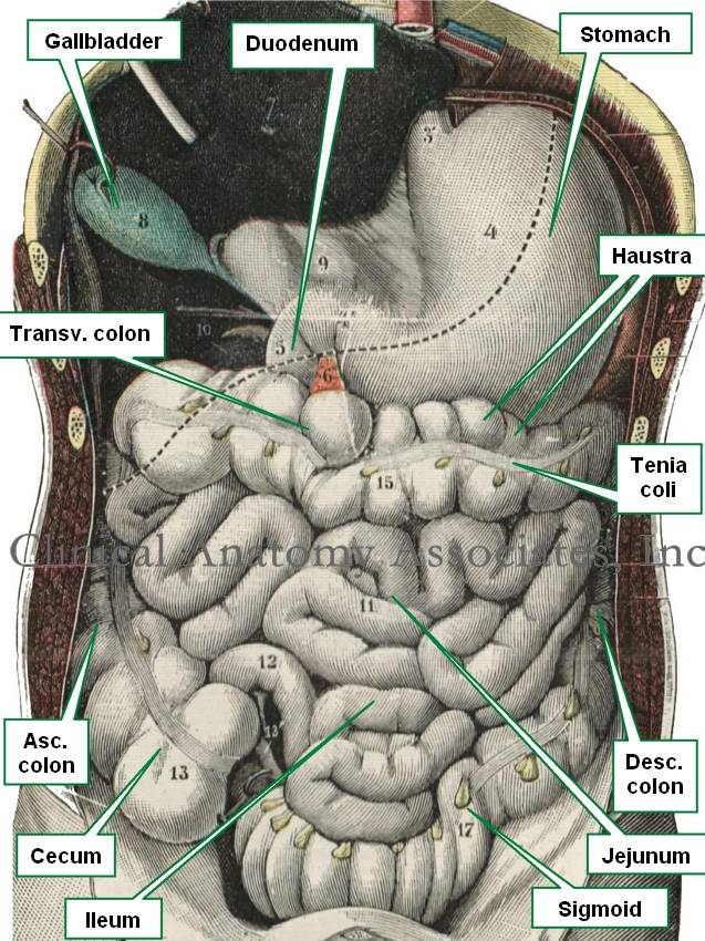
In anatomy, the word [haustrum] is used to refer to a sacculation or outpouching of the colon caused by the constant tonus contraction of the longitudinal muscle fibers found in the tenia coli, as well as the distribution of circular fibers. See accompanying image. Click the image for a larger depiction.

Since the small intestine is not necessarily always small in relation to the colon (because of food content, intestinal gases, or pathology), the location of the organ and the presence of haustra, as well as the presence of tenia coli and appendices epiploica, is used to recognize the organ as colon.

The term can also be used as an adjective and the colon can be said to be "haustrated".

The word seems to have been used for the first time by Albrecht Von Haller (1708 - 1777) a Swiss anatomist, botanist, and physiologist.

Sources:
1. "Dorlands's Illustrated Medical Dictionary" 26th Ed. W.B. Saunders 1994
2. "The origin of Medical Terms" Skinner, AH, 1970
3. "Tratado de Anatomia Humana" Testut et Latarjet 8 Ed. 1931 Salvat Editores, Spain
Image modified from the original from Testut and Latajet, 1931

Abdominal contents (Testut Latarjet 1931)
Image modified from the original from Testut and Latajet, 1931

Back to MTD Main Page Subscribe to MTD

-algia

The medical suffix [-algia] originates from the Greek [algos /algein] meaning "pain". The term is used in many medical words. Applications of this root term include:

• Arthralgia: The root term [-arthr-] means "a joint", or "an articulation".Pain in a of a joint
Cephalalgia: The root term [-cephal] means "head". A headache
Myalgia: The root term [-my(o)] means "muscle". Muscle pain
Otalgia: The root term [-ot-] means "ear". An earache
Neuralgia: The root term [-neur-] means "nerve". Pain of nerve origin
Prosopalgia: From the Greek [pros?pon]shaping of a joint

Back to MTD Main Page Subscribe to MTD

Ileum

The ileum is an intraperitoneal organ, it is the third portion of the small intestine and part of the digestive tract, found distal to the duodenum and jejunum. The ileum is about 6-8 feet in lenght.

There is no clear anatomical boundary between the jejunum and ileum, as they blend smoothly one into the other. There are several gross changes from jejunum to ileum, one of them being that the complexity of the mesenteric arterial arches increases from proximal to distal.

The term [ileum] has a Greek origin from [ειλεός] meanings "twisted", referring to the "twisted" look of this third portion of the small intestine. When the term was first used Galen did not separate or differentiate jejunum from ileum. He called  both the [intestinum tenuis] or "thin or delicate intestine". This term is still used for the smaller arterial and venous branches of the small intestine, the [vasa intestinii tenuis].

It seems that the term "ileum" or "ileus" was originally used to denote the distal portion of the intestine that got sick or "colicky", separating it from the jejunum wich was usually found empty or devoid of food.

The terminal ileum ends at the ileocolic or ileocecal valve, where the small intestine empties its contents into the large intestine or colon.

Abdominal contents (Testut Latarjet 1931)

Sources:
1. "Clinically Oriented Anatomy" Moore, KL. 3r Ed. Williams & Wilkins 1992
2. "The origin of Medical Terms" Skinner, AH, 1970
3. "Tratado de Anatomia Humana" Testut et Latarjet 8 Ed. 1931 Salvat Editores, Spain
Image modified from the original from Testut and Latajet, 1931. Click in the image for a larger depiction
Back to MTD Main Page Subscribe to MTD