Medical Terminology Daily (MTD) is a blog sponsored by Clinical Anatomy Associates, Inc. as a service to the medical community. We post anatomical, medical or surgical terms, their meaning and usage, as well as biographical notes on anatomists, surgeons, and researchers through the ages. Be warned that some of the images used depict human anatomical specimens.

You are welcome to submit questions and suggestions using our "Contact Us" form. The information on this blog follows the terms on our "Privacy and Security Statement" and cannot be construed as medical guidance or instructions for treatment.


We have 168 guests online


A Moment in History

Jean-Louis Petit

Jean Louis Petit
(1674 – 1750)

French surgeon and anatomist, Jean Louis Petit was born in Paris in on March 13, 1674.  His family rented an apartment at his house to Alexis Littre (1658 – 1726), a French anatomist. Petit became an apprentice of Littre at seven years of age, helping him in the dissections for his lectures and at an early age became the assistant in charge of the anatomic amphitheater.

Because of Petit’s dedication to anatomy and medicine, in 1690 at the age of sixteen, became a disciple of a famous Paris surgeon, Castel.

In 1692, Petit entered the French army and performed surgery in two military campaigns. By 1693 he started delivering lectures and was accepted as a great surgeon, being invited to the most difficult operations.  In 1700 he was appointed Chief Surgeon of the Military School in Paris and in the same year he received the degree of Master of Surgery from the Faculty of Paris.

In 1715 he was made a member of the Royal Academy of Sciences and an honorary member of the Royal Society of London. He was appointed by the King as the first Director General of the Royal Academy of Surgery when it was founded in 1731.

Petit’s written works are of historical importance.  “Traite des Maladies des Os” ( A Treatise on Bone Diseases);  “Traite des Maladies Chirurgicales et des Operation” (A Treatise on Surgical Diseases and their Operations” This last book was published posthumously in 1774. He also published a monograph on hemorrhage, another on lachrymal fistula, and others.

He was one of the first to perform choIecystotomy and mastoidotomy. His original tourniquet design for amputations saved many in the battlefield and the design of the same surgical instrument today has not changed much since its invention by him.

His name is remembered in the lumbar triangle, also called the "triangle of Petit", and the abdominal hernia that can ensue through that area of weakness, the lumbar hernia or "Petit's hernia".

Sources:
1. “Jean Louis Petit – A Sketch of his Life, Character, and Writings” Hayne, AP San Fran Western Lancet 1875 4: 446-454
2. “Oeuvres compl?tes de Jean-Louis Petit” 1837 Imprimerie de F. Chapoulaud
3. Extraits de l'eloge de Jean-Louis Petit Ius dans Ia seance publique de I' Academie royale de chirurgie du 26 mai 1750” Louis A. Chirurgie 2001: 126 : 475- 81


 "Clinical Anatomy Associates, Inc., and the contributors of "Medical Terminology Daily" wish to thank all individuals who donate their bodies and tissues for the advancement of education and research”.

Click here for more information


abebooks banner

bookplateink.com


Manubrium

UPDATED: The word [manubrium] is Latin and mean "handle", referring to the area where a person holds an instrument or device. To exemplify this, in Spanish the vernacular use of the word [manubrio] refers to the handles of bicycle or even the steering wheel of a car. 

In anatomy, the term is used with the same meaning. In the malleus, a hammer-like ossicle of the middle ear, the manubrium is the handle-like extension of the bone that attaches to the tympanic membrane.

In the case of the sternum, the [manubrium sterni] is the superior portion bound by the sternal angle (of Louis) inferiorly.  The use of the word manubrium can be explained because in early anatomy, the sternum was known by the Latin term [gladius] referring to the similarity of the sternum to the short sword of the gladiators. The area where you hold the sword is the handle, ergo, manubrium.

The manubrium has a superior and median notch called the "suprasternal notch" or the "jugular notch". It is important because in the case of a mediastinoscopy, the incision is made just superior to this landmark. The manubrium articulates superolaterally with the clavicle and inferolaterally with the superior aspect of the cartilage of the second rib. The rest of the rib cartilage articulates with the body of the sternum.

Image property of:CAA.Inc.. Artist: Mark J. Zuptich

Sternal angle - Angle of Luis
Click on the image for a larger version.
MTD Main Page Subscribe to MTD

Induration

The word [induration] arises from the Latin words induratio, meaning "thick or hard" and indurare, meaning "hardening".

It refers to a pathological hardening of tissues caused by tumoration or edema, increase of fibrous or connective tissue, or other causes. It is a good, descriptive term when stating a patient's symptoms. The term has been in use in English since the 14th century.

Note: The links to Google Translate include an icon that will allow you to hear the pronunciation of the word.

Back to MTD Main Page Subscribe to MTD

It's our 20th year anniversary!!!

20 year Anniversary

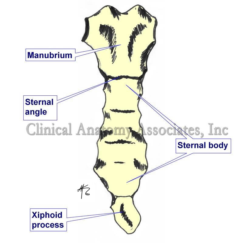
At the beginning of 1998, Clinical Anatomy Associates, Inc. was formed as an Ohio Corporation. Our mission is to deliver industry relevant, cutting-edge Training, Marketing, and R&D services that will enable our clients to gain a competitive advantage. Over the past two decades, Clinical Anatomy Associates, Inc. has become the go-to R&D resource for feasibility studies that require cadaver studies and anatomical research. We are also a preferred training solution for Sales Representatives, Distributors, Engineers, Clinicians, and Marketing Managers in the areas of Medical Terminology, Clinical Anatomy, and Surgical Procedures. Our expertise allows us to deliver training in a variety of medical and anatomical topics.

In 2012 Dr. Efrain A. Miranda, CEO of Clinical Anatomy Associates started "Medical Terminology Daily" (MTD), a website/blog as a service to the medical community, medical students, and the medical industry. MTD posts medical or surgical terms, its meaning and usage, as well as biographical notes on anatomists, surgeons, and researchers through the ages. These posts are also shared on Facebook to a group of followers.

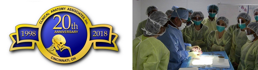
20 year anniversary for Clinical Anatomy Associates, Inc. and 6 years for Medical Terminology Daily! Help us congratulate our staff and specially the contributors and friends of Medical Terminology Daily.

Our thanks to all our customers, friends, and contributors for an amazing 20 years!!! Looking forward to more!!

Back to MTD Main Page Subscribe to MTD

Happy New Year 2018!

The staff at Clinical Anatomy Associates, Inc. wishes a Happy and Prosperous New Year 2018 to the readers, subscribers, contributors, and friends of Medical Terminology Daily.

This year we are looking at bringing in new contributors, new articles, and updating our website to help our visitors even more

HAPPY NEW YEAR 2018!!

MTD Main Page Subscribe to MTD

Snowman sign

The “snowman sign” is a particular image on a chest X-Ray image, which is seen in anomalous pulmonary venous drainage and coarctation of the aorta which causes a Total Anomalous Pulmonary Venous Return (TAPVR).

This abnormality occurs when the pulmonary veins fail to drain into the left atrium and instead form an aberrant connection with some others cardiovascular structures. Such abnormalities account for approximately 2% of cardiac malformations.

There are four types of TAPVR; type 1 is the most common (and the one that creates the snowman sign). In this case the pulmonary veins terminate at the supracardiac level, emptying into the right atrium by way of an anomalous pulmonary venous drainage into the superior vena cava (SVC), and the left brachiocephalic vein (by way of a vertical vein). The confluence of these veins dilates the right brachiocephalic vein, which appears as a dilated vessel on the right of the upper mediastinal edge. When seen in an AP Chest X-Ray, the TAPVAR type 1, resembles a snowman; the dilated vertical vein on the left, the right brachiocephalic vein superiorly, and the SVC on the right form the head of the snowman, the body is formed by the enlarged right atrium.

Article written by: Prof. Claudio R. Molina, MsC

Snowman sign
Snowman sign.
Click on the image for a larger depiction

Sources:
1. Emma C. Ferguson, Rajesh Krishnamurthy, and Sandra A. A. Oldham. (2007) Classic Imaging Signs of Congenital Cardiovascular Abnormalities. RadioGraphics 27:5, 1323-1334.
2. Somerville, J., & Grech, V. (2009). The chest x-ray in congenital heart disease 1. Total anomalous pulmonary venous drainage and coarctation of the aorta. Images in Paediatric Cardiology, 11(1), 7–9.

Back to MTD Main Page Subscribe to MTD

Kabourophobia

Kabourophobia is the fear of crabs and lobsters.

The etymology of the word [kabourophobia] comes from the Greek word [καβουρης] (pronounced “kavouris”), meaning [crab], and the suffix [-phobia], also from the Greek, arises from the word [φοβία] (pronounced “fovía”)

Kabourophobia is an extremely rare phobia, but it was brought to the public’s attention when a modern pop singer stated that she was afraid of crabs. Also, a prank (maybe acted) was shown on video on the internet with a man surrounded by lobsters.

Kabourophobia is very specific, and it can also be a part of a wider phobia called ostraconophobia, which is the fear of crustaceans, adding shrimp, oysters, clams, crabs, lobsters, etc.

 Liocarcinus vernalis © Hans Hillewaert  via Wikimedia Commons

Click on the image for a larger version. 

An interesting point is that the word [crab] in Greek has another acception, that is the word [Καρκίνος] (pronounced “karkinos”), which is the root for the medical term [cancer].

We thank Jackie Miranda-Klein for her contribution suggesting this word.

MTD Main Page Subscribe to MTD