Medical Terminology Daily (MTD) is a blog sponsored by Clinical Anatomy Associates, Inc. as a service to the medical community. We post anatomical, medical or surgical terms, their meaning and usage, as well as biographical notes on anatomists, surgeons, and researchers through the ages. Be warned that some of the images used depict human anatomical specimens.

You are welcome to submit questions and suggestions using our "Contact Us" form. The information on this blog follows the terms on our "Privacy and Security Statement" and cannot be construed as medical guidance or instructions for treatment.


We have 237 guests online


A Moment in History

Jean-Louis Petit

Jean Louis Petit
(1674 – 1750)

French surgeon and anatomist, Jean Louis Petit was born in Paris in on March 13, 1674.  His family rented an apartment at his house to Alexis Littre (1658 – 1726), a French anatomist. Petit became an apprentice of Littre at seven years of age, helping him in the dissections for his lectures and at an early age became the assistant in charge of the anatomic amphitheater.

Because of Petit’s dedication to anatomy and medicine, in 1690 at the age of sixteen, became a disciple of a famous Paris surgeon, Castel.

In 1692, Petit entered the French army and performed surgery in two military campaigns. By 1693 he started delivering lectures and was accepted as a great surgeon, being invited to the most difficult operations.  In 1700 he was appointed Chief Surgeon of the Military School in Paris and in the same year he received the degree of Master of Surgery from the Faculty of Paris.

In 1715 he was made a member of the Royal Academy of Sciences and an honorary member of the Royal Society of London. He was appointed by the King as the first Director General of the Royal Academy of Surgery when it was founded in 1731.

Petit’s written works are of historical importance.  “Traite des Maladies des Os” ( A Treatise on Bone Diseases);  “Traite des Maladies Chirurgicales et des Operation” (A Treatise on Surgical Diseases and their Operations” This last book was published posthumously in 1774. He also published a monograph on hemorrhage, another on lachrymal fistula, and others.

He was one of the first to perform choIecystotomy and mastoidotomy. His original tourniquet design for amputations saved many in the battlefield and the design of the same surgical instrument today has not changed much since its invention by him.

His name is remembered in the lumbar triangle, also called the "triangle of Petit", and the abdominal hernia that can ensue through that area of weakness, the lumbar hernia or "Petit's hernia".

Sources:
1. “Jean Louis Petit – A Sketch of his Life, Character, and Writings” Hayne, AP San Fran Western Lancet 1875 4: 446-454
2. “Oeuvres compl?tes de Jean-Louis Petit” 1837 Imprimerie de F. Chapoulaud
3. Extraits de l'eloge de Jean-Louis Petit Ius dans Ia seance publique de I' Academie royale de chirurgie du 26 mai 1750” Louis A. Chirurgie 2001: 126 : 475- 81


 "Clinical Anatomy Associates, Inc., and the contributors of "Medical Terminology Daily" wish to thank all individuals who donate their bodies and tissues for the advancement of education and research”.

Click here for more information


abebooks banner

bookplateink.com


Did Andreas Vesalius really die from scurvy? (1)


Did Andreas Vesalius really die from scurvy on the island of Zakynthos in 1564?
Evidence does not support this theory.

NOTE:  The following article authored by Theo Dirix, and Dr. Rudi Coninx, is a rebuttal of Pavlos Plessas' theory that Andreas Vesalius indeed died from scurvy. The original article entitled "Powerful indications that Vesalius died from scurvy" by Pavlos Plessas was presented in a meeting at the island of Zakynthos in 2014.


Andreas Vesalius, the father of modern anatomy, was a famous man in his time. Born Andries van Wesel in 1514 in Brussels, he became the most important authority on anatomy of his time, daring even to correct the teachings of Galen, the ultimate reference on anatomy since the ancient times. Author of the most famous book on anatomy, the seven-volume "De Humani Corporis Fabrica, Libri Septem" , professor of surgery and anatomy in Padua at age 23 in 1537, imperial physician to the court of King Charles V and personal physician of the emperor of the Spanish Empire, King Philip II, Andreas Vesalius’ accomplishments are multifold. Yet, surprisingly, there are lots of things we do not know about him: how did he look like, for example, but also –and that is the topic of this paper- how did he die, what did he die from, and where exactly is he buried?

There have been more items related to Vesalius’ life that were wrongly transmitted through time, but recent investigations have put to rest a number of “disputed” facts that were erroneously reported and commonly accepted. They often made into the general history books or even into Wikipedia. The two most glaring ones were

1. That Andreas Vesalius had to leave the Imperial Court in Spain because the Inquisition was investigating him for allegedly doing an autopsy on a person that was not yet dead.

Theo Dirix and Dr. Rudi Coninx, authors of this article
Dr. Rudi Coninx, and Theo Dirix, authors of this article

• We now know that Andreas Vesalius did not leave the Imperial Court for his pilgrimage to Jerusalem under pressure of the Inquisition, as is often alleged [1], but that he left on his own accord and with the full support of the Emperor who provided him with introduction letters to Spanish embassies abroad. We thought the Vesalius and the inquisition myth was laid to rest in 1928 [2], but has to be refuted again and again to this day, it seems [3].

2. That Andreas Vesalius was shipwrecked, and presumably drowned

• Vesalius did not die shipwrecked, as it was still alleged by Wikipedia recently [4] (but now corrected. His ship arrived safely at the harbour of the island of Venetian owned island of Zante (now Zakinthos, Greece) and Andreas Vesalius went ashore there.

What do we know for sure about the last days of Andreas Vesalius? 

• We know that Andreas Vesalius undertook a pilgrimage to Jerusalem in early March 1564, arriving in Venice late April, and that he sailed from Venice via Cyprus to Jerusalem in early May. He only stayed for a short time in Jerusalem, probably one month. He then traveled from Jerusalem through the Sinai desert to Alexandria, Egypt and boarded a Venetian ship there in September 1564, heading for Venice. 

• We know that the return journey was eventful. The ship was initially driven off course and then stuck without winds for over forty days which lead to food and water shortages on the ship. Most passengers fell ill, and many died. Their bodies were thrown overboard. This caused great anxiety to Andreas Vesalius who feared being thrown overboard unceremoniously in case he died. Vesalius became ill himself and lay sick in the hold with nobody taking care of him. (Account of Reinert Soleander, quoted by Biesbrouk et al.) [5].

• We also know that he stepped off the ship on the Venetian island of Zante (now Zakynthos in Greece) on October 15, 1564, sick, weak and frail, that he slowly walked towards the gates of the city where he collapsed and fell dead.

These are the facts that can be attested, and are not in dispute. What remains disputed is: did this 50 year old man die from scurvy, as is alleged?

In this article we will look at the arguments advanced in favour of the scurvy theory and see if there is any evidence to support these arguments.

Most arguments in favour of the scurvy theory have been proposed by Mr. Pavlos Plessas at the 2014 Vesalius Continuum meeting in Zakynthos, Greece and posted online on his blog (and here) [5]:

• “Vesalius stayed for six months in an area where Vitamin C was not easily found.
• Vesalius’s travels and sightseeing during the summer months means that some of his body’s Vitamin C reserves were lost through perspiration.
• Over the duration of his pilgrimage this could have exceeded 20% of his initial reserves. He then stayed an additional forty days at sea becoming vitamin C deficient.
• He reports Vesalius as immobile, depressed, with phobic and paranoid behavior,
• These can have sudden death syndrome upon restart of physical activity, which would also explain Vesalius' sudden death at the doors of the port of Zakynthos [7]..


Article continued here: Did Andreas Vesalius really die from scurvy? (2)
Sources:

1. https://circulatingnow.nlm.nih.gov/2014/10/15/the-death-of-andreas-vesalius/ accessed 27.12.2016
2. Matheson Cullen, G. Vesalius and the inquisition myth. Lancet, January 14, 1928, p 105-6.
3. Dirix Th. In search of Andreas Vesalius. The quest for the lost grave. Lannoo, 2014.
4. https://en.wikipedia.org/wiki/Andreas_Vesalius accessed on January 21, 2016
5. Biesbrouck M, Goddeeris Th, Steeno O. The last months of Andreas Vesalius. A coda. In Vesalius, Acta Internationalia Historiae Medicinae. 2012, 18 (No 2), 70-75.
6. Plessas P. http://www.parathemata.com/2014/09/pavlos-plessas-powerful-indications.html 2014. Accessed January 21, 2016.
7. http://www.clinicalanatomy.com/andreas-vesalius
Aa https://ods.od.nih.gov/factsheets/VitaminC-HealthProfessional/
8. Fain, O. La Revue de Médecine Interne, 2004; vol 25, Issue 12, 872-880.
9. Hodges RE, Hood J, Canham HE, Sauberlich HE, Baker EM. Clinical manifestations of ascorbic acid deficiency in man. Am J Clin Nutr 1971;24:432-43.
x. Hirschman JV, Raugi GJ. Adult scurvy. Journal of American Academy of Dermatology, 1999, 41; No 6, 895-909.
xx. Bartley W, Krebs HA, O’Brien JRP. Vitamin C requirement of human adults. Medical Research Council Special Report Series No 280. London: Her Majesty’s Stationary Office; 1953. P 1-179. Quoted in Hirschmann et al.
xxx Hodges RE, Baker EM, Hood J, Saueberlich HE, March SC. Experimental scurvy in man. American Journal of Clinical Nutrition. 1969;22:535-48.
10. Harrisons Principles of Internal Medicine, 1998; p 484-85
11. Leung FW, Guze PA: Adult scurvy. Annals of Emergency medicine; 1981; 10:652-655
12. Bennet M, Coninx R. The mystery of the wooden leg: vitamin C deficiency in East African prisons. Tropical Doctor, 2005; 35: 81-84.
13. Carpenter K. The history if scurvy and vitamin C.Cambridge University Press 1986, p29.
11. Bown S R. 4he Age of Scurvy. How a surgeon, a mariner and a gentleman helped Britain win the battle of Trafalgar. Summersdale, 2003, p 96-99.
15. Lind J. A treatise of the scurvy. Containing an inquiry into the nature, causes and cure of that disease. Together with a critical and chronological view of what has been published on the subject. Edinburgh: Sands, Murray and Cochran: 1753.
16. Kinsman RA, Hood J: Some behavioural effects of ascorbic acid deficiency. The American Journal of Clinical Nutrition, 1971, 455-464.
17. Biesbrouck M, Goddeeris Th, Steeno O: ‘Post Mortem’ Andreae Vesalii (1514 – 1564). Deel II. Het graf van Andreas Vesalius op Zakynthos. A. Vesalius, nr.4 December 2015. [in Dutch].
18. Bruce M Rothschild. Scurvy imaging. http://emedicine.medscape.com/article/413463-overview

MTD Main Page Subscribe to MTD

Glossary of Terms for Thoracic Imaging

One of the current challenges in medical sciences is to achieve a common and universal language that allows establishing, promoting the understanding and communication between people of different cultures and nationalities.

The use of anatomical and radiological terms in the chest is not exempt from this theme. An adequate terminology allows describing findings and unifying concepts that facilitate communication, teaching, research, in brief, the transmission of knowledge.

From the radiological point of view, ignorance of appropriate terminology creates confusion and may eventually lead to inaccuracies and misdiagnoses. Therefore, reaching consensus in terminology is an attempt to minimize the effects of language vices, which are unfortunately passed on to future generations.

On the other hand, in recent years, technical advances in radiology have improved the capture, recording and storage of images, which in turn has involved the need for a new language to explain new concepts.

Fleischner
Felix Fleischner, MD
Within this context, the Fleischner Society, an international and multidisciplinary medical society for thoracic radiology, was founded in 1969 in memory of Dr. Félix Fleischner. Among the objectives of this society is the publication of formal statements that have been adopted for several years, as standards in the field of chest radiology, generating an important point of discussion and consensus around this issue.

Thus, as in 1971 a proposal for initial terminology was made, which was consolidated in 1984 and 1996 with the publications of the Fleischner Society glossary for Chest X-rays and Tomographies respectively. In 2008 (Radiology 2008; 246: 697-722) this glossary is finally updated using new terms, modifying or eliminating some of those that have become obsolete and others in which their meaning has changed.

Sources:
1. Austin JHM, Müller NL, Friedman PJ. Glossary of Terms for CT of the Lungs: Recommendations of the Nomenclature Committee of The Fleischner Society. Radiology 1996; 200:327-30.
2. Fraser RS, Müller NL, Colman N, Pare PD. Diagnosis of diseases of the Chest. 4th ed. Philadelphia: WB Saunders Company, 1999.
3. Tuddenham WJ. Glossary of Terms for Thoracic Radiology: Recommendations of the Nomenclature Committee of The Fleischner Society. Am J Roentgenol 1984; 143:509-17.
4. Souza Jr AS, Araújo Neto CA, Jasinovodolinki D,Marchiori E, Kavakama J, Irion KL et al. Terminologia para a Descrição de Tomografia Computadorizada do Tórax (Sugestões Iniciais para um Consenso Brasileiro). Radiol Bras 2002; 35:125-8.
5.David M. Hansell, Alexander A. Bankier, Heber MacMahon, Theresa C. McLoud, Nestor L. Müller, and Jacques Remy Fleischner Society: Glossary of Terms for Thoracic Imaging Radiology 2008 246:3, 697-722


This article belongs to the series "Glossary of Terms for Thoracic Imaging" by Prof C. Uribe, MsC. This series is based on the "Glossary of Terms for Thoracic Imaging" by the Fleischner Society
Back to MTD Main Page Subscribe to MTD

Johann Gottfried Zinn

This article is part of the series "A Moment in History" where we honor those who have contributed to the growth of medical knowledge in the areas of anatomy, medicine, surgery, and medical research.

Johann Gottfried Zinn

Johann Gottfried Zinn (1727–1759) anatomist and botanist, was born on December 6, 1727 in the city of Ansbach, Germany. He started his medical studies in his native city, becoming later a student of Dr.  Albrecht von Hallers at the University of Göttingen, and received his MD in 1749.

He left for Berlin to continue his studies but came back shortly thereafter. He became a professor of anatomy at the University of Göttingen and in 1753 he also became the director of the botanical garden in the same city.

He is known for his anatomical treatise on the anatomy of the human eye: “Descriptio anatomica oculi humani iconibus illustrata”. Because of this, his name has become an eponym in the “Zonule of Zinn”, a ring of strands that forms a fibrous band connecting the ciliary body with the capsule of the lens of the eye. Zonule of Zinn is sometimes referred to as the suspensory ligaments of the lens, or the “ligament of Zinn”. His name is also attached to the anular ring tendon found in the posterior aspect of the eye, the "anular tendon of Zinn". This ring serves as attachment for all the extraocular muscles of the eye and the optic nerve passes through the center of the ring.

Carol Linné (Carolus Linneaus) named a genus of flowers in the family Asteraceae known vernacularly today as “Zinnia” in his honor. Hover your cursor over his portrait to see the flower.

The chapter on orbital anatomy of his anatomy book, taken from the second edition in 1780, has been translated and the first of three parts is published in an issue of “Strabismus”

His book "Catalogus Plantarum Horti Academici Et Agri" can be read online here.

His life was short, dying at the early age of 32, but his name lives on in the name of a beautiful flower.

Sources:
1. “Johann Gottfried Zinn" Simonz, HJ Strabismus – 2004, Vol. 12, No.
2, p. 125 2. "Anatomical Description of the Human Eye" Zinn, JG Strabismus, 13:45–52, 2005
Images: Public Domain by Wikipedia Commons. 1. Own work I_am Jin, and H. Wilhem Dietz


Update to the "A Moment in History" Series

This article is part of the series "A Moment in History" where we honor those who have contributed to the growth of medical knowledge in the areas of anatomy, medicine, surgery, and medical research.

Václav Treitz
Vaclav Treitz

As part of the redesign of this website we added a sidebar called "A Moment in History". The objective is to create a series of articles to honor those individuals who have contributed to the growth of medical knowledge in the areas of anatomy, medicine, surgery, and medical research. Later in the development of the series we became aware of other individuals who have contributed in different ways, but still added their life work to the advancement of medical knowledge, as is the case of Marcia Croker Noyes (1869-1946).

Who would not be moved by the work of Allesandra Gilliani (1307-1326), who is probably the first woman dissector in the history of Human Anatomy, with a tragic short life and a love story.

We also decided to add to this series Moments in History that have left a mark on health care, such as "The First Use of Anesthesia in Surgery", or the story of how many individuals and unknown, anonymous children helped to rid the Americas from the scourge of smallpox, in "The Balmis Expedition", 

Another line of articles in this series are those that honor individuals who have used anatomical and surgical knowledge to further other areas of human knowledge, such as that of Juan Vucetich, who used the anatomical differences in fingerprints to create the science of dactiloscopy.

Yet another line of articles are those that are more personal and dear to the contributors of "Medical Terminology Daily", such as "The Ephraim McDowell House of Museum", or  "Interesting Discovery in and Ex-Libris".

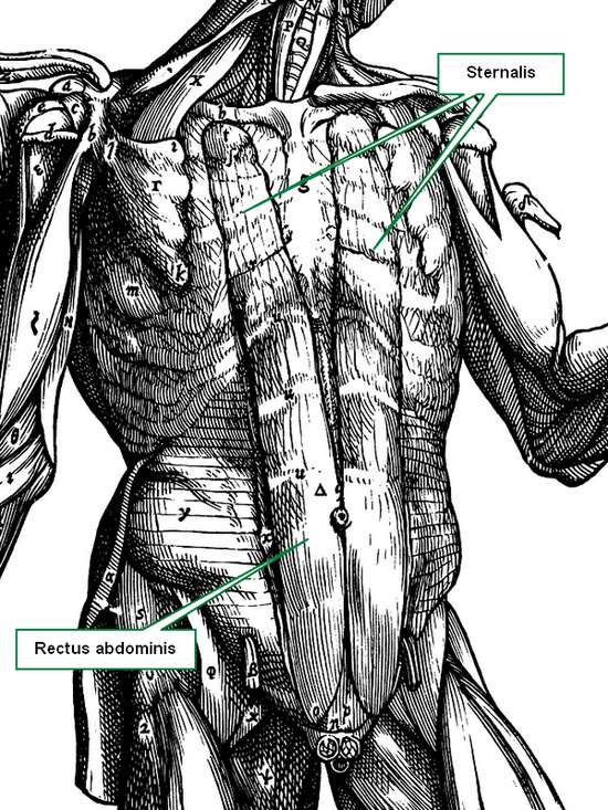
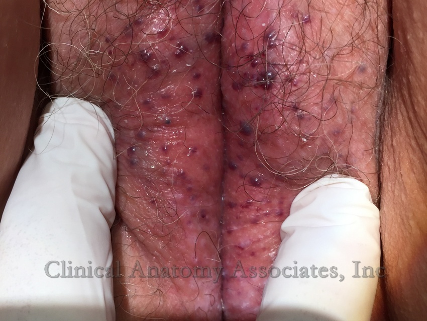
Recently, I had to work in the Wangensteen Historical Library researching rare and antique medical books. The highlight of this work was to be able to read books by authors whose names are attached as eponyms to anatomical landmarks (Ligament of Treitz, Hesselbach's Triangle), pathologies (Koplik's spots), surgical procedures (Billroth I and II), medical maneuvers (Heimlich maneuver), and surgical instruments (Finochietto retractor). Of course, the names given here are but a small sample of what has been written to date.

As of today this series is now searchable, all you have to do is type "A Moment in History" in our search page, click on the "A Moment in History" link at the top of the sidebar, or click here

The image in this article is that of Dr. Vaclav Treitz. His eponymically named Ligament of Treitz is the most read article in this blog.

Original image, public domain, courtesy of Wikipedia.org.


Sternalis muscle

The Sternalis muscle has been nominated as a supernumerary anatomical variation; the highest prevalence is in China (23.5%) and the lowest in Chile (0.87%).

The muscle is a wide band of muscular tissue located in the anterior thoracic wall, where fibers travel near-parallel to the sternum, inferior to the clavicles, aterior to the pectoralis major and the pectoral fascia. It has received many names: rectus sternalis, parasternal, episternal, superficial abdominal rectus, among other names. To avoid confusion, a true sternalis muscle should have the following characteristics:

(1) be localized between the subcutaneous tissue of the anterior thoracic region and fascia pectoralis;
(2) originate superiorly from the sternum or infraclavicular region;
(3) be inserted inferiorly in the ribs, rib cartilage, external oblique muscle aponeurosis and sheath of the rectus abdominis muscle;
(4) its innervation must come from the anterior root of the intercostal nerves. Another possible innervation from medial and lateral pectoral nerves has been mentioned.

Even though the Sternalis muscle does not appear to have a significant function, it is important to bear it in mind when evaluating mammograms, because one could misdiagnose it as a mammary cancer, as exposed by many authors. It is crucial to be familiar with this muscle to avoid confusion with pathology, such as extra-abdominal desmoid tumors, diabetic mastopathy, abscesses, hematomas, fat necrosis, and others; its unilateral presence may cause breast or chest asymmetry.

Sternalis muscle (Andreas Vesalius 1543)
Note: The image in this article depicts this variation and is from the 1543 " De Humanis Corporis Fabrica, Libri Septem " by Andreas Vesalius.

Sources:
1. Jelev L., Georgiev G., Surchev L. (2001) The sternalis muscle in the Bulgarian population: classification of sternales. J. Anat. 199: 359-363. 
2. Bradley F.M., Hoover H.C., Hulka C.A., Whitman G.J., McCarthy K.A., Hall D.A., Moore R., Kopans D.B. (1996) The sternalis muscle: an unusual normal finding seen on mammography. AJR. Am. J. Roentgenol. 166: 33-36. 
3. Scott-Conner C.E.H., Al-Jurf A.S. (2002) The sternalis muscle. Clin. Anat. 15: 67-69. 
4. Shiotani M., Higuchi T., Yoshimura N., Kiguchi T., Takahashi N., Maeda H., Aoyama H. (2012) The sternalis muscle: radiologic findings on MDCT. Jpn. J. Radiol. 30: 729-734.


Back to MTD Main Page Subscribe to MTD

Vulvar hemangiomata

UPDATED: This article presents a case of vulvar hemangiomata. In superficial or surface anatomy, [vulva] is the anatomical name given to the external female genitalia.

The medical term [hemangioma] is formed by two root terms and a suffix. The root term [hem-] arises from the Greek word [αίμα] (a?ma) meaning "blood", the second root term [-angi-] .from the Greek term [αγγείο] (angeio), meaning "vessel” and the suffix [-oma] (ωμα), also Greek, meaning "mass", "growth”, or "tumor". The plural derivative word [hemangiomata] uses the suffix [-omata] which is the plural form of [-oma].

Vulvar hemangiomata are multiple blood growths or tumors on a female’s external genitalia. In most cases, hemangiomata are idiopathic.

Vulvar hemangiomata
Image property of: CAA.Inc. 
WARNING: Depicts female external genitalia.
Vulvar hemangiomata can be capillary (arterial) or cavernous (venous). They are benign vascular tumors which can be congenital, meaning the patient is born with them, or acquired, meaning that they appear later in life. In both cases they are usually asymptomatic. Patients may become concerned over aesthetics or because of bleeding due to trauma to the lesions. In rare cases, these hemangiomas can be large, requiring excision.

They are usually kept under observation, evaluating their progress (evolution or involution). If they increase in number or become symptomatic, intervention includes the use of sclerosing agents, destruction by cryotherapy or laser therapy, or surgical excision.

Personal note: My thanks to the patient who voluntarily and anonymously provided this image to further learning and knowledge on this pathology. Dr. Miranda

MTD Main Page Subscribe to MTD