Medical Terminology Daily (MTD) is a blog sponsored by Clinical Anatomy Associates, Inc. as a service to the medical community. We post anatomical, medical or surgical terms, their meaning and usage, as well as biographical notes on anatomists, surgeons, and researchers through the ages. Be warned that some of the images used depict human anatomical specimens.

You are welcome to submit questions and suggestions using our "Contact Us" form. The information on this blog follows the terms on our "Privacy and Security Statement" and cannot be construed as medical guidance or instructions for treatment.


We have 127 guests online


A Moment in History

Jean-Louis Petit

Jean Louis Petit
(1674 – 1750)

French surgeon and anatomist, Jean Louis Petit was born in Paris in on March 13, 1674.  His family rented an apartment at his house to Alexis Littre (1658 – 1726), a French anatomist. Petit became an apprentice of Littre at seven years of age, helping him in the dissections for his lectures and at an early age became the assistant in charge of the anatomic amphitheater.

Because of Petit’s dedication to anatomy and medicine, in 1690 at the age of sixteen, became a disciple of a famous Paris surgeon, Castel.

In 1692, Petit entered the French army and performed surgery in two military campaigns. By 1693 he started delivering lectures and was accepted as a great surgeon, being invited to the most difficult operations.  In 1700 he was appointed Chief Surgeon of the Military School in Paris and in the same year he received the degree of Master of Surgery from the Faculty of Paris.

In 1715 he was made a member of the Royal Academy of Sciences and an honorary member of the Royal Society of London. He was appointed by the King as the first Director General of the Royal Academy of Surgery when it was founded in 1731.

Petit’s written works are of historical importance.  “Traite des Maladies des Os” ( A Treatise on Bone Diseases);  “Traite des Maladies Chirurgicales et des Operation” (A Treatise on Surgical Diseases and their Operations” This last book was published posthumously in 1774. He also published a monograph on hemorrhage, another on lachrymal fistula, and others.

He was one of the first to perform choIecystotomy and mastoidotomy. His original tourniquet design for amputations saved many in the battlefield and the design of the same surgical instrument today has not changed much since its invention by him.

His name is remembered in the lumbar triangle, also called the "triangle of Petit", and the abdominal hernia that can ensue through that area of weakness, the lumbar hernia or "Petit's hernia".

Sources:
1. “Jean Louis Petit – A Sketch of his Life, Character, and Writings” Hayne, AP San Fran Western Lancet 1875 4: 446-454
2. “Oeuvres compl?tes de Jean-Louis Petit” 1837 Imprimerie de F. Chapoulaud
3. Extraits de l'eloge de Jean-Louis Petit Ius dans Ia seance publique de I' Academie royale de chirurgie du 26 mai 1750” Louis A. Chirurgie 2001: 126 : 475- 81


 "Clinical Anatomy Associates, Inc., and the contributors of "Medical Terminology Daily" wish to thank all individuals who donate their bodies and tissues for the advancement of education and research”.

Click here for more information


abebooks banner

bookplateink.com


Vasa vasorum

[UPDATED] The term [vasa vasorum] comes from the Latin [vasa] or [vas] meaning "a vessel", as in a container. Vasa vasorum means "the vessels of the vessels"

In arteries and veins, only the inner layer of the vessel, the endothelium can receive nutrition and oxygen from the blood that is carried by the vessel. Arteries and veins have their own blood supply network, their own arteries and veins. These are the vasa vasorum.

The image shows the internal structure of an artery with its three layers, tunica intima (named after Xavier Bichat), tunica media, and tunica adventitia, as well as the vasa vasorum. Click on the image for a larger depiction. 

 Vasa vasorum. Artwork in black and white by Michiaki Akashi, M.D.
NOTE: My personal thanks to Michiaki Akashi, M.D. for allowing us to use his artwork in this article. Dr. Akashi works as a surgeon and pathologist in the Saga Prefectural Hospital Koseikan in Saga, Japan. Dr. Miranda
Back to MTD Main Page Subscribe to MTD

Obturator foramen

The [obturator foramen] is a bilateral and roughly triangular opening in the lower portion of the pelvis bound by the following bones: ilium, ischium, and pubic bone. The obturator foramen is closed off by the obturator membrane, a though, tendinous membrane that leaves a small superior opening (obturator canal) that allows passageway of the obturator nerve, obturator artery, and obturator vein. These neurovascular structures provide nerve and blood supply to the anterior compartment of the thigh.

The obturator membrane serves as a point of attachment to the external obturator and internal obturator muscles.

The obturator foramen is important for transobturator sling surgery used in cases of urinary incontinency. 

Image property of: CAA.Inc. Artist: M. Zuptich

Obturator foramen 

Back to MTD Main Page Subscribe to MTD

Lambda

The  word [lambda] is Greek and is represented by the symbols [Λ] and [λ]. Because of the inverted "V" shape and inverted "Y" shape of these symbols, it was used by Galen of Pergamon (129AD - 200AD) to denote the junction of the sagital suture (interparietal articulation) and the occipitoparietal suture, also known as the "lamboid" suture. The lambda can also be described as the point of junction of the occipital bone with the parietal bones.

Although Galen named this junction originally, it was Vesalius (1514- 1564) who brought this name into modern anatomy. Later, Peter Paul Broca (1824- 1880) used this landmark as a craniometric point.

Click on the accompanying image of a posterior view of the cranium for a larger depiction.

Image in public domain, modified from Toldt's "Atlas of Human Anatomy", 1903.

Cranium, posterior view. (Toldt's Anatomy Atlas)
Back to MTD Main Page Back to MTD Main Page

Leonardo Botallus


This article is part of the series "A Moment in History" where we honor those who have contributed to the growth of medical knowledge in the areas of anatomy, medicine, surgery, and medical research.To search all the articles in this series, click here.

Leonardo Botallus (c.1530- ??) Italian anatomist and physician,Leonardo Botallus (also known as Botalli, Botallo, or Botal) was born circa 1530 in the region of Piedmont. Botallus studied medicine at the italian university of Pavia, where he was a student under Gabrielle Fallopius.  Botallus graduated circa 1553.  He was an avid advocate of bloodletting, causing him to direct his anatomical studies towards the subject of the vascular system. Although he did not discover the foramen ovale and the ductus arteriosus, he mentions these structures by those names in both his posthumous  publications " De via sanguinis a dextro in sinistro cordis ventriculum" (1640) and "Opera Omnia Medica et Chirurgica" (1660)

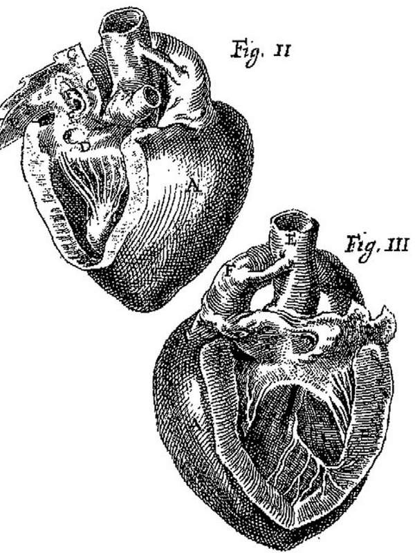
In France Botallus became physician to King Charles IX. Not much more is known about Botallus, and  history fails to record his date and place of death, as well as his image. The image depicted here is from his 1660 publication. Click on the image for a larger depiction.

Botallus' name is eponymically remembered in the following structures:

Image from Botallus '<em>Opera Omnia Anatomica et Chirurgica'</em>

Foramen of Botallus: The foramen ovale, an opening found in the fetus in the region of the fossa ovalis that closes upon birth
Duct of Botallus: A communicating vessel between the left pulmonary artery and the proximal region of the descending aorta, part of fetal circulation, also known as the ductus arteriosus
Ligament of Botallus: The closed ductus arteriosus in the adult

Sources:
1. "History of medicine; a correlative text, arranged according to subjects" Mettler, C Ch. 1947 The Blakiston Co 
2. "Stedmans Medical Eponyms" Forbis, P; Bartolucci, SL. Williams & Wilkins 1998
3. "The origin of Medical Terms" Skinner, AH, 1970
4. "The First Closure of the Persistent Ductus Arteriosus" Alexi-Meskishvili,V; B?ttcher, W. Ann Thorac Surg 2010;90:349 –56
NOTE: There is no known image of Botallus. Skinner's "Origin of Medical Terms" shows one, but we could not confirm the origin of the image. If you know or have one, let us know

Back to MTD Main Page Subscribe to MTD

Basivertebral Foramen

[UPDATED] The basivertebral foramen is an opening  in the posterior aspect of each vertebra (click on the picture for a larger image) which allows the exit of the basivertebral veins. These foramina can be single (as seen in the image) or multiple. The basivertebral veins represent a communication that allows drainage of the vertebral body venous sinuses into the extensive complex venous network of the internal venous plexuses that surrounds the spinal cord.

Photography by D.M.Klein

Thoracic vertebra, posterior view
Back to MTD Main Page Back to MTD Main Page